


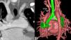
A two-year-old girl was admitted to the hospital following the incidental discovery of a heart murmur three days previously. A grade 3/6 continuous murmur was audible near the left sternal border at the second intercostal space. Chest X-ray suggested increased bilateral pulmonary blood flow and cardiomegaly. Transthoracic echocardiography revealed the right aortic arch (RAA), patent ductus arteriosus (PDA) and the left subclavian artery (LSA) originating from the pulmonary artery. Computed tomography angiography (CTA) images showed a bilateral ductous arteriosus with concomitant combinations of RAA and isolated LSA. Right-sided PDA connected from the descending aorta to the right pulmonary artery and left-sided PDA connected the LSA to the left pulmonary artery with meandering and narrowing (Figure 1A and B). The patient underwent bilateral PDA ligation and LSA reconstruction under cardiopulmonary bypass. Postoperative recovery was uneventful, and the patient was discharged after seven days.

(A) Maximal intensity projection (MIP) of computed tomography angiography (CTA) images with a coronal image shows bilateral patent ductus arteriosus with an isolated left subclavian artery originating from the left pulmonary artery. (B) Three-dimensional volume rendering (3D VR) of CTA image shows a bilateral arteriosus with concomitant combinations of RAA and isolated LSA. Right-sided PDA connected from the descending aorta to the right pulmonary artery and left-sided PDA connected the LSA to the left pulmonary artery with meandering and narrowing. RIA, right innominate artery; LSA, left subclavian artery; LPDA, left patent ductus arteriosus; RPDA, right-sided patent ductus arteriosus.
Double or bilateral ductus arteriosus (BDA) is a rare anomaly. This anomaly is frequently associated with a right aortic arch and other anomalies, including double aortic arch, discontinuous central PAs, anomalous subclavian artery, or isolation of brachiocephalic vessels.1 The embryology of the development is based on the double arch theory. BDA is formed when the distal ends of the sixth pair of primitive arches on the left and right sides have not regressed.2 BDA with an isolated LSA associated with RAA can result in a vascular ring, if it compresses the trachea and esophagus, causing related symptoms.3 In addition, it may manifest as a subclavian or pulmonary steal, vertebrobasilar insufficiency and congestive heart failure.4
It is essential to recognize this abnormality associated with cardiac anomalies. Understanding the clinical features, embryology, and imaging findings is necessary for an accurate diagnosis and clinical treatment. Due to the extreme rarity of our case, there is no consensus for management. Treatment of BDA depends upon the other cardiac or non-cardiac abnormalities; many patients require complex surgical correction and cardiac catheterization with BDA stenting.5
Ethical approvalThe Institutional Review Board of Wuhan Aisa Heart Hospital approved this study and the patient's informed consent was waived (IRB number: SOP-LLWYH-025-03-R1).
Conflicts of interestThe authors have no conflicts of interest to declare.
