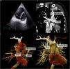
A 54-year-old man was referred for assessment of right chamber dilatation. He had been previously diagnosed with left superior vena cava (SVC) persistence at the age of 30 but had no other relevant medical history. A transthoracic echocardiogram confirmed severe right chamber dilatation with diastolic flattening of interventricular septum (Figure 1A). Right systolic ventricular function was normal. There was no tricuspid regurgitation. Abnormal continuous flow originating in the inferior region of the aortic arc and directed toward the transducer was observed from the suprasternal window. There was no coronary sinus dilatation. The injection of agitated saline into the left antecubital vein did not show opacification of the coronary sinus nor the presence of bubbles in the left cavities.
Transthoracic echocardiography parasternal short axis view showing severe dilatation of the right ventricle (panel A). Cardiac computed tomography showing both right and left superior pulmonary veins draining into the left brachiocephalic vein through a vertical vein. An accessory left superior pulmonary vein is observed (panels B and C). Reconstructed three-dimensional cardiac computed tomography imaging showing the vertical vein draining into the left brachiocephalic vein (panel D). LBV: left brachiocephalic vein; LSPV: left superior pulmonary vein; RSPV: right superior pulmonary vein; SVC: superior vena cava.
A cardiac computed tomography demonstrated a bilateral partial anomalous pulmonary venous drainage (PAPVD). Both right and left superior pulmonary veins drained through a vertical vein into the left brachiocephalic vein (Figure 1B–D). The inferior pulmonary veins had normal anatomy and drained into the left atrium. The pulmonary trunk was dilated (39 mm×32 mm). No interatrial or interventricular communications were observed.
Bilateral PAPVD of both superior pulmonary veins is rare and this anatomy is seldom described in the literature.1 Misdiagnosis of a left-sided SVC from a vertical vein associated with anomalous pulmonary venous drainage into the brachiocephalic vein is possible. Nevertheless, this differentiation should be carefully considered, as important clinical and therapeutic implications may arise.2
FundingThe authors declare that no funds, grants, or other support were received during the preparation of this manuscript.
Conflict of interestThe authors report no conflicts of interest.


