Journal Information
Vol. 32. Issue 2.
Pages 169-171 (February 2013)
Vol. 32. Issue 2.
Pages 169-171 (February 2013)
Image in cardiology
Open Access
Usefulness of cardiac computed tomography in planning and evaluating alcohol septal ablation
Utilidade da tomografia computorizada cardíaca no planeamento e avaliação do resultado da ablação septal por álcool
Visits
7733
Ana Faustinoa,
Corresponding author
anacatarina.faustino@gmail.com

Corresponding author.
, Nuno Ferreirab, Nuno Bettencourtb, Mónica Carvalhob, Daniel Leiteb, Vasco Gamab
a Serviço de Cardiologia, Centro Hospitalar e Universitário de Coimbra, Coimbra, Portugal
b Serviço de Cardiologia, Centro Hospitalar de Vila Nova de Gaia/Espinho, Gaia, Portugal
This item has received

Under a Creative Commons license
Article information
Full Text
Bibliography
Download PDF
Statistics
Figures (4)
Show moreShow less
Full Text

Alcohol septal ablation (ASA) is an option in the treatment of obstructive hypertrophic cardiomyopathy refractory to medical therapy.1–3 Cardiac computed tomography (CT) is useful in the selection of patients, as it can simultaneously assess the coronary anatomy and its spatial relations with the myocardium, which is essential in determining the feasibility of ASA and selecting the appropriate septal artery. It can also evaluate the success of the procedure, by assessing the obstruction decreasing and the extent of fibrotic tissue (without additional contrast if performed immediately following ASA), at low dose radiation exposure.1–3

The authors present images illustrating the value of CT, comparing it with the other imaging modalities normally used in this context.

Figure 1 compares images of left ventricular outflow tract (LVOT) obstruction by echocardiography (A) and CT (B), the latter also identifying a septal artery (arrow).

Figure 1.

(A): Two-dimensional transthoracic echocardiogram in parasternal long-axis view in diastole; (B): the same view by multiplanar reconstruction (MPR) cardiac CT, 12 mm thickness, in diastole.

(0.17MB).

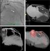
Figure 2 shows the anatomy of the septal arteries by invasive coronary angiography (A) and CT (B–D), showing the relationship between the septal arteries (arrow) and the ventricular chamber (C) and the hypertrophied myocardium (B and D).

Figure 2.

(A): Invasive coronary angiography in right anterior oblique view; (B): maximum intensity projection (MIP) CT coronary angiography, 12 mm thickness; (C) and (D): CT coronary angiography, three-dimensional reconstruction.

(0.53MB).

Images of the LVOT (Figure 3) by echocardiography (A) and CT (B) following ASA confirm the success of the procedure.

Figure 3.

(A): Two-dimensional transthoracic echocardiogram in parasternal long-axis view in systole; (B): cardiac CT (MPR), 12 mm thickness, 3-chamber view, in systole (25% of the cardiac cycle).

(0.19MB).

Figure 4 compares late enhancement images by magnetic resonance imaging (A) and CT (B) following ASA, showing areas of myocardial fibrosis (arrows) and microvascular lesion induced by alcohol ablation (triangle).

Figure 4.

(A): Cardiac magnetic resonance (phase-sensitive inversion recovery), late enhancement study in short-axis view; (B): cardiac CT (MPR), 12 mm thickness, in the same view.

(0.23MB).

Cardiac CT is a non-invasive technique that can be used to plan ASA, assess the outcome and predict its long-term success.

Ethical disclosuresProtection of human and animal subjects

The authors declare that no experiments were performed on humans or animals for this study.

Confidentiality of data

The authors declare that they have followed the protocols of their work center on the publication of patient data and that all the patients included in the study received sufficient information and gave their written informed consent to participate in the study.

Right to privacy and informed consent

The authors declare that no patient data appear in this article.

Conflicts of interest

The authors have no conflicts of interest to declare.

References
[1]
J.F. Deux, J. Potet, P. Lim, et al.
Is multidetector computed tomography a suitable alternative to MR imaging for the assessment of myocardial necrosis after alcohol septal ablation.
[2]
R. Mitsutake, S. Miura, H. Sako, et al.
Usefulness of multi-detector row computed tomography for the management of percutaneous transluminal septal myocardial ablation in patient with hypertrophic obstructive cardiomyopathy.
Int J Cardiol, 129 (2008), pp. e61-e63
[3]
S. Okayama, S. Uemura, T. Soeda, et al.
Role of cardiac computed tomography in planning and evaluating percutaneous transluminal septal myocardial ablation for hypertrophic obstructive cardiomyopathy.
J Cardiovasc Comput Tomogr, 4 (2010), pp. 62-65

Please cite this article as: Faustino, A; Utilidade da tomografia computorizada cardíaca no planeamento e avaliação do resultado da ablação septal por álcool. Rev Port Cardiol 2013. http://dx.doi.org/10.1016/j.repc.2012.07.009

Copyright © 2012. Sociedade Portuguesa de Cardiologia
Download PDF
Idiomas
Revista Portuguesa de Cardiologia (English edition)
Article options
Tools
en pt

Are you a health professional able to prescribe or dispense drugs?

Você é um profissional de saúde habilitado a prescrever ou dispensar medicamentos

By checking that you are a health professional, you are stating that you are aware and accept that the Portuguese Journal of Cardiology (RPC) is the Data Controller that processes the personal information of users of its website, with its registered office at Campo Grande, n.º 28, 13.º, 1700-093 Lisbon, telephone 217 970 685 and 217 817 630, fax 217 931 095, and email revista@spc.pt. I declare for all purposes that the information provided herein is accurate and correct.