Congenital coronary artery anomalies affect therapeutic options when associated with congenital heart disease.
Echocardiography is the first-line exam for diagnosis and management of patients with congenital heart disease, but is less useful for anatomical assessment of the great vessels and the coronary arteries. Cardiac catheterization has a higher risk of complications due to its invasive nature, and since it is a two-dimensional technique, the anatomical information it provides is limited.
In the last two decades, computed tomography (CT) and magnetic resonance imaging have become important non-invasive diagnostic tools that provide more accurate anatomical information than any other imaging modality.
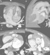
The authors describe the CT findings in a 29-month-old boy referred for cardiac CT angiography to assess complex congenital heart disease. The study was performed on a 128-slice prospectively ECG-gated dual-source Siemens Definition Flash system and showed mesocardia with abdominal/atrial situs inversus (Figure 1), atrioventricular and ventriculoarterial discordance with a subpulmonary ventricular septal defect, and a single coronary artery (Figure 2). Effective radiation dose was 1.0 mSv (dose-length product 25 mGy.cm).
Mesocardia and situs inversus. (A) Topogram; (B) multiplanar reconstruction, coronal view; (C) axial image of the abdomen; (D) three-dimensional volume rendering showing mesocardia, morphological right bronchus in left position, morphological left bronchus in right position, liver on the left, stomach and spleen on the right, and anterior right aorta and posterior left pulmonary artery. Ao: aorta: B: spleen; E: stomach; F: liver; P: pulmonary artery.
Atrioventricular and ventriculoarterial discordance with a single coronary artery. Axial (A) and multiplanar (B, C and D) reconstructions showing atrioventricular and ventriculoarterial discordance, with anterior right aorta and posterior left pulmonary artery, infundibular septal defect with subarterial communication (asterisk), and anomalous origin of the right coronary artery in the left coronary sinus, coursing between the aorta and the pulmonary artery. AD: right atrium; AE: left atrium; Ao: aorta; CD: right coronary artery; Cx: circumflex artery; DA: anterior descending artery; P: pulmonary artery; VD: right ventricle; VE: left ventricle.
The patient was considered unsuitable for surgery due to the high surgical risk arising from the single coronary artery. This case illustrates the value of cardiac CT angiography for detailed non-invasive anatomical assessment of intra- and extracardiac structures, and was decisive in the therapeutic decision-making process.
Conflicts of interestThe authors have no conflicts of interest to declare.
Please cite this article as: Duarte R, Sampaio MA, Morais H, et al. Artéria coronária única com mesocardia, situs inversus, discordância aurículo-ventrículo e ventrículo-arterial 2013;32:837–838.


