


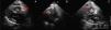
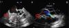
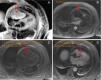
A 65-year-old man with a history of thrombocytopenia currently under investigation, hypertension and diabetes, was hospitalized with decompensated heart failure, mainly right-sided. Transthoracic echocardiography (Figure 1) showed preserved biventricular function and a relatively immobile heterogeneous mass, 2.5 cm in diameter, in the right ventricle (RV), that was difficult to characterize because of poor echocardiographic image quality and patient discomfort. Transesophageal echocardiography (Figure 2), interrupted due to dyspnea, revealed that the mass was adhering to the lateral wall of the right atrium (RA) and extended to the tricuspid valve (causing obstruction), the RV and the pericardium. Provisional diagnoses were thrombus or tumor, its heterogeneous appearance and infiltrative nature suggesting the latter. Thoracic-abdominal-pelvic computed tomography was accordingly performed to investigate a possible occult tumor, but this showed no alterations other than bilateral pleural effusion and a large cardiac mass occupying the right chambers and obliterating the external RA wall. Cardiac magnetic resonance imaging (Figure 3) showed a neoplasm adhering to the lateral RA wall and protruding into the RV, suggestive of an intracardiac tumor.
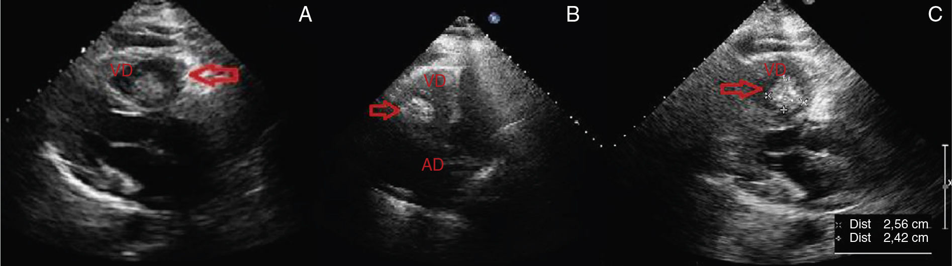
Transthoracic echocardiography 24 hours after admission showing a relatively immobile heterogeneous mass (arrow) in the right ventricle and slight pericardial effusion. (A) Mass in long-axis parasternal view; (B) mass in right ventricle in apical 4-chamber view; (C) parasternal long-axis view showing measurements of the mass. AD: right atrium; VD: right ventricle.
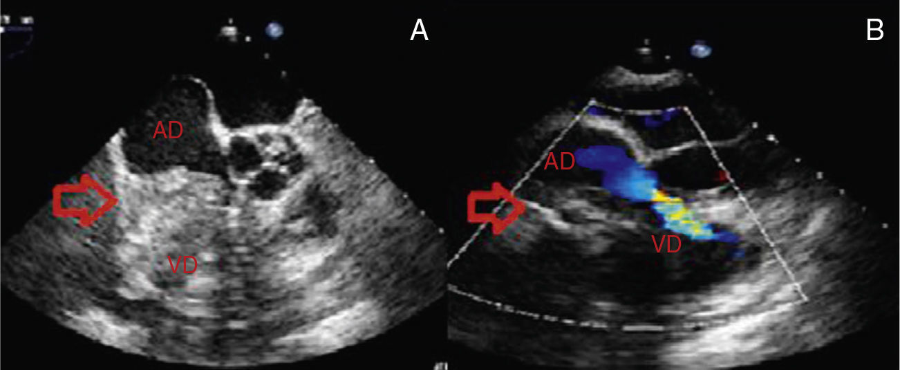
Transesophageal echocardiography to investigate the intracardiac mass (arrow). (A) Relatively immobile heterogeneous mass with both hypo- and hyperechogenic areas, adhering to the lateral wall of the right atrium and extending to the tricuspid valve, right ventricle and pericardium; (B) mass obstructing flow in the right chambers. AD: right atrium; VD: right ventricle.
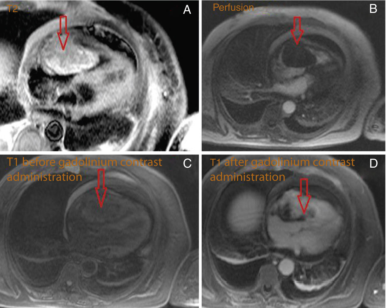
Cardiac magnetic resonance imaging showing an intracavitary mass (arrow) adhering to the lateral wall of the right atrium, measuring 56 mm×87 mm×47 mm, protruding above the atrial appendage, through the tricuspid valve, into the right ventricle and below the atrial wall. (A) Relatively homogenous high signal intensity in T2 and equal signal intensity compared to the myocardium in T1 (C and D); predominantly central enhancement throughout the perfusion study (B), with areas of peripheral hypoperfusion. Slight pericardial effusion can be seen.
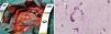
The patient underwent surgical resection of the mass (Figure 4A) and implantation of a mechanical tricuspid valve, but he died 16 hours after the operation following refractory shock. Histological study of the surgical specimen (Figure 4B) showed it to be an osteosarcoma, rarely found in the heart as either a primary or secondary tumor. In this case it was considered to be primary, given the lack of evidence of tumors in other locations (osteosarcoma usually affects the long bones), even though primary tumors are more common in the left chambers, unlike secondary tumors, which are more frequent in the right heart.

Macroscopic (A) and microscopic (B) views of the tumor. (A) Tumor (T), solid, occupying most of the right atrium; (B) histological study of the surgical specimen showing mesenchymal, epithelioid (E) and osteoid (O) proliferation, with focal mineralization (M), suggesting osteosarcoma (hematoxylin–eosin stain). AD: right atrium; VD: right ventricle.
The authors have no conflicts of interest to declare.
Ethical disclosuresProtection of human and animal subjectsThe authors declare that the procedures followed were in accordance with the regulations of the relevant clinical research ethics committee and with those of the Code of Ethics of the World Medical Association (Declaration of Helsinki).
Confidentiality of dataThe authors declare that they have followed the protocols of their work center on the publication of patient data and that all the patients included in the study received sufficient information and gave their written informed consent to participate in the study.
Right to privacy and informed consentThe authors have obtained the written informed consent of the patients or subjects mentioned in the article. The corresponding author is in possession of this document.
Please cite this article as: Damásio AF, Vasconcelos J, Pós de Mina V, et al. Insuficiência cardíaca direita – qual a etiologia? Rev Port Cardiol. 2014;33:191–193.
