Renal artery aneurysms are a rare cause of secondary hypertension. Endovascular treatment with a polytetrafluoroethylene (PTFE)-coated stent can exclude aneurysms and treat hypertension. We report the case of a 23-year-old man with hypertension diagnosed three years earlier and in whom renal angiography revealed three aneurysms involving the right renal artery. A covered stent was implanted, resulting in successful exclusion of the aneurysm. Ten months after the procedure the patient is asymptomatic and with normal blood pressure without antihypertensive therapy.
Os aneurismas da artéria renal representam uma causa rara de hipertensão secundária. O tratamento endovascular com stent revestido por politetrafluoretileno (PTFE) consiste numa abordagem que permite excluir os aneurismas e tratar a hipertensão. Apresentamos o caso de um jovem de 23 anos, com o diagnóstico de hipertensão há três anos e cuja angiografia renal mostrou três aneurismas na dependência da artéria renal direita. Procedeu-se à implantação de um stent revestido por PTFE com a exclusão bem sucedida dos aneurismas. Dez meses após o procedimento o doente está assintomático e com valores normais de tensão arterial sem terapêutica anti hipertensora.
Hypertension is the most prevalent risk factor for cardiovascular disease and is frequently poorly controlled. Despite the long list of causes for hypertension, in about 90% of cases the etiology is unknown. However, the prevalence of secondary hypertension is increasing with improved methods of diagnosis.1 Renal artery aneurysms (RAAs) are found at 0.3–0.7% of autopsies and in up to 1% of renal arteriographic procedures.2 Fibromuscular dysplasia, degenerative aneurysms, vasculitis and trauma are the most frequent causes. These aneurysms have a female predilection. RAAs are associated with hypertension in up to 73% of cases.3 Hypotheses on the pathophysiological basis of hypertension include microembolization from the aneurysm, coexisting renal artery stenosis, compression or kinking of the renal artery or its branches, and turbulent flow.3
Management decisions should be based on patient age and gender, anticipated pregnancy, severity of hypertension and anatomic features of the aneurysm.4 Pregnant women, female patients of childbearing age, and patients with evidence of embolization are candidates for surgical or endovascular intervention.5 Other causes for such interventions are symptomatic aneurysms (flank pain, hypertension, hematuria), rapidly expanding aneurysms and those larger than 2cm.6 The choice of treatment of RAAs is determined by the anatomic location of the aneurysm.5 Although stent grafts and stent-plus-coil embolization techniques are successful for most simple RAAs,7 complex aneurysms beyond the bifurcation of the main renal artery, or those involving major arterial branches, may require extracorporal arterial reconstruction followed by autotransplantation.6
Case reportWe describe the case of a 23-year-old Caucasian man diagnosed with hypertension three years previously and no other relevant personal or family history.
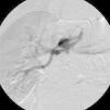
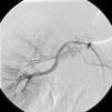
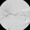
Non-invasive 24-hour blood pressure monitoring revealed stage I hypertension (mean daytime blood pressure 156/87mmHg). Hormonal and imaging studies were also performed for etiologic diagnosis of his hypertension, as well as a captopril test, which was positive for renovascular hypertension. Following this result, renal angiography was performed, which revealed, bilaterally, two renal arteries, and in addition, to the right, depending on the superior artery, three saccular aneurysms 14mm, 6mm and 3.5mm in size. The largest aneurysm appeared to compress the lower polar artery (Figure 1). The aneurysms were treated with placement of a polytetrafluoroethylene (PTFE)-coated stent, in order to prevent expansion and rupture of the aneurysms and to treat the hypertension. Digital subtraction angiography was performed using a right femoral approach. A 65cm 4F sheath was introduced, and the right renal artery was engaged with a 4F Cobra catheter and a 0.014-inch hydrophilic guidewire. Aneurysm morphology was assessed using conventional angiography and flat-panel computed tomography. Stent diameter and length were determined from a three-dimensional flat-panel rotational angiography data set. After administration of 5000 IU of heparin, the aneurysm was crossed with the guidewire and catheter. The wire was exchanged through the same catheter with a 0.014-inch guidewire. Finally, a 6×22-mm covered Atrium stent was deployed, bridging the aneurysm and covering the artery, resulting in successful exclusion of the aneurysms (Figures 2 and 3). The patient was admitted the day before the procedure and was discharged the day after, medicated with 100mg aspirin and 75mg clopidogrel/day, as dual antiplatelet therapy. Ten months after the procedure the patient was asymptomatic, with normal blood pressure and without antihypertensive therapy.
The development and wide dissemination of non-invasive imaging techniques has led to the diagnosis of an increasing number of new cases of secondary hypertension. Cases formerly designated as essential hypertension now have a specific diagnosis, often with the possibility of resolution, avoiding the effects of high blood pressure and the chronic use of antihypertensive therapy. The clinical relevance of incidentally discovered RAAs remains the subject of debate, with uncertainty regarding the threshold for intervention.8 However, there is a consensus that repair should be performed in pregnant women or those of childbearing age, in cases with evidence of embolization,5 in symptomatic aneurysms (flank pain, hypertension, hematuria), and in rapidly expanding aneurysms and those larger than 2cm.6 The case described concerns a young male diagnosed with hypertension three years earlier and under antihypertensive therapy. English et al. reported the surgical outcome of 62 patients treated for RAAs, 89% of whom had hypertension; most of these (75%) experienced beneficial blood pressure response in a mean follow-up of 48 months after RAA repair, including 21% who became normotensive off all medications.4 Other surgical series report similar rates of improvement in hypertension following RAA treatment in conjunction with fewer antihypertensive medications.3,9 Due to the clinical repercussions of aneurysms in this patient and the expected benefits of the intervention, there was no doubt regarding the need for treatment, leading to a multidisciplinary discussion on the best form of approach. Three general approaches have been described for RAA treatment: (1) surgery with either in situ aneurysmectomy and bypass with an autologous conduit, or nephrectomy9; (2) transcatheter embolization5; or (3) endovascular exclusion by stent graft.10,11 The angiographic pattern of RAAs and their feeding artery or arteries help to determine the optimal method of treatment. Three main types of RAAs are described: saccular RAAs arising from either the main renal artery or a large segmental branch (type 1), which are the most common; fusiform aneurysms (type 2); and intralobar aneurysms (type 3), which arise from small segmental arteries that supply a limited portion of the renal parenchyma.10 Type 1 RAAs can be successfully treated with stent graft implantation; type 2 are best treated by a surgical approach, while type 3 may be treated with catheter-directed embolization using microcoils.10 However, morbidity and long recovery periods persist in cases of open surgical repair, and aortorenal bypass occlusions and unplanned nephrectomies occur even in the largest series. Additionally, some patients may not be candidates for surgical repair because of comorbidities that preclude surgery or because of complex aneurysm morphology or location.8 Recently, laparoscopic surgery has been proposed as a minimally invasive alternative to open surgical RAA repair.12 The percutaneous treatment of RAAs with covered stents, increasingly accepted as safe and effective, also offers several advantages compared to traditional surgical therapy due to its minimally invasive nature; it may be a safe therapeutic alternative to surgery in cases with an appropriate artery caliber proximal and distal to the aneurysm, and with the aneurysm neck not located close to a branching point of the renal artery. The treatment of RAAs with covered stents has some limitations because of the inflexibility of the devices, preventing application in small and tortuous vessels, and due to the risk of thrombosis and stenosis.8 Different embolic techniques (such as selective coil embolization), remodeling techniques (including balloon and stent-assisted coiling), and embolization with liquid embolic agents (such as glue or onyx) exist and in selected patients represent the first-line treatment option for RAAs. However, in these techniques also it is sometimes necessary to perform a parent-vessel occlusion to treat the aneurysm with some degree of renal parenchyma compromise.8
In the case presented, a multidisciplinary consensus led to a conservative approach with implantation of a PTFE-coated stent in which the aneurysms involved a large-caliber vessel, the aneurysms were easily accessible, and there were no collateral vessels of significant size in the area of deployment of the stent. On the other hand, coil embolization of an important vessel may result in non-target embolization or coil migration or delayed recanalization of the aneurysm. The risk of occlusion reinforces the importance of maintaining dual antiplatelet therapy.
The procedure was uneventful and the final images show the successful exclusion of the aneurysms. Ten months after the procedure, the absence of symptoms and normalization of blood pressure show the success of the intervention.
ConclusionThe endovascular approach has demonstrated safety and effectiveness in treating selected patients with renovascular hypertension. Due to low procedure-related mortality and morbidity, percutaneous renal interventions have been incorporated into common practice despite the lack of evidence from randomized studies to support their efficacy over the long term. The cases described in the literature indicate good prognosis in hypertension control and exclusion of aneurysms, and the present case demonstrates the efficacy and safety of the percutaneous approach in treating RAAs.
Conflicts of interestThe authors have no conflicts of interest to declare.


