Mid-aortic syndrome is an uncommon condition characterized by constriction of the distal thoracic and/or abdominal aorta and its branches.
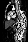
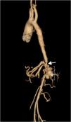
A 43-year-old male patient with a history of neurofibromatosis type 1 (NF-1) was referred to our hospital for resistant arterial hypertension. At physical examination he presented asymmetric elevated blood pressure in the extremities, higher in the upper limbs. An echocardiogram was performed and showed moderate left ventricular hypertrophy, without signs of coarctation of the aorta. As a part of workup for hypertension, computed tomography (CT) was performed, which showed an abnormal aorta with severe narrowing and a saccular aneurysm at the level of the renal arteries (Figures 1 and 2). There was extensive collateral blood flow through hypertrophic lumbar, epigastric and mesenteric arteries and the left renal artery showed subtotal stenosis at its origin, causing atrophy of the left kidney (Figure 3). Additional angiographic study enabled a final diagnosis. There were also subcutaneous and retroperitoneal neurofibromas.
Mid-aortic syndrome usually presents with arterial hypertension and is commonly diagnosed in children or young adults. It can be associated with Williams syndrome, Takayasu arteritis, NF-1, Alagille syndrome and Moyamoya disease. Surgery is the primary treatment when it is associated with renovascular hypertension and visceral artery stenosis. Endovascular therapy can be performed in patients with discrete aortic stenosis; however, due to inherent arterial wall anomalies, vascular complications increase after percutaneous procedures, thus making them poor options. Our patient refused surgical procedures and has been treated with medical therapy. Prognosis is generally good, but the syndrome may have high morbidity and mortality if left untreated.
Ethical disclosuresProtection of human and animal subjectsThe authors declare that no experiments were performed on humans or animals for this study.
Confidentiality of dataThe authors declare that no patient data appear in this article.
Right to privacy and informed consentThe authors declare that no patient data appear in this article.
Conflicts of interestThe authors have no conflicts of interest to declare.


