Journal Information
Vol. 35. Issue 3.
Pages 191-193 (March 2016)
Share
Share
Download PDF
More article options
Vol. 35. Issue 3.
Pages 191-193 (March 2016)
Image in Cardiology
Open Access
Inferior wall acute myocardial infarction or something else?
Enfarte agudo do miocárdio da parede inferior ou algo mais?
Visits
4454
Leandro Rassi
Corresponding author
leandrorassi6@gmail.com

Corresponding author.
, Rita Rodrigues, Catarina Sá, Tatiana Duarte, Ricardo Santos, Rui Caria
São Bernardo Hospital, Department of Cardiology, Setúbal, Portugal
This item has received

Under a Creative Commons license
Article information
Full Text
Download PDF
Statistics
Figures (5)
Show moreShow less
Additional material (6)
Full Text

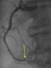
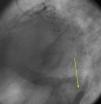
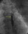
The authors present the case of an 83-year-old male with a history of hypertension, appendectomy and cervical spine surgery who presented at the emergency department with epigastric pain and dyspnea at rest for six hours. The physical examination was compatible with refractory acute pulmonary edema requiring endotracheal intubation and ventilation. The electrocardiogram showed sinus rhythm with ST-segment elevation in the inferior leads. The emergent coronary angiography revealed three-vessel disease with distal occlusion of the right coronary artery (Figure 1, Videos 1 and 2). Primary angioplasty was performed successfully (Figure 2, Video 3). During the procedure a contrast retention image was observed apparently synchronous with the cardiac/respiratory cycle (Figure 3, Video 4). The first possibility that came to mind was an aortic dissection, which was excluded by an anteroposterior projection, which showed that the image was in the midline (Figure 4, Videos 5 and 6). A chest computed tomography scan was then requested to rule out the presence of a fistula (esophageal or bronchial), which revealed that the image corresponded to retention of contrast in the medullary canal from myelography performed in the 1980s (Figure 5A and B).

Figure 1.

Distal occlusion of the right coronary artery (arrow).

(0.05MB).
Figure 2.

Successful primary angioplasty of the right coronary artery.

(0.04MB).
Figure 3.

Contrast retention image on coronary angiography (arrow).

(0.06MB).
Figure 4.

Coronary angiography, anteroposterior projection, showing the contrast retention image in the midline (arrow).

(0.05MB).
Figure 5.

Chest computed tomography scan showing contrast retention in the medullary canal (arrows).

(0.07MB).

Myelography is still used in the study of patients with suspected atrophic or degenerative changes in the spinal cord. Current soluble contrast agents have less acute and chronic neurotoxicity than previously used agents and do not need to be removed. In the case presented it was not possible to obtain information as to why the contrast agent was not removed after the procedure.

Ethical disclosuresProtection of human and animal subjects

The authors declare that no experiments were performed on humans or animals for this study.

Confidentiality of data

The authors declare that no patient data appear in this article.

Right to privacy and informed consent

The authors declare that no patient data appear in this article.

Conflicts of interest

The authors have no conflicts of interest to declare.

Appendix A
Supplementary material

(0.74MB)
(1.01MB)
(0.55MB)
(0.59MB)
(0.56MB)
(0.57MB)

Copyright © 2016. Sociedade Portuguesa de Cardiologia
Download PDF
Idiomas
Revista Portuguesa de Cardiologia (English edition)
Article options
Tools
Supplemental materials
en pt

Are you a health professional able to prescribe or dispense drugs?

Você é um profissional de saúde habilitado a prescrever ou dispensar medicamentos

By checking that you are a health professional, you are stating that you are aware and accept that the Portuguese Journal of Cardiology (RPC) is the Data Controller that processes the personal information of users of its website, with its registered office at Campo Grande, n.º 28, 13.º, 1700-093 Lisbon, telephone 217 970 685 and 217 817 630, fax 217 931 095, and email revista@spc.pt. I declare for all purposes that the information provided herein is accurate and correct.