


The value of cardiac computed tomography (CT) in the assessment of coronary anatomy is well established.1 This technique also enables visualization of other cardiac structures, with constantly improving resolution, and incidental findings are thus relatively common.
The authors present three cases of intracardiac findings during cardiac CT.
Case 1A 20-year-old man complaining of chest pain was referred for cardiac CT to exclude coronary artery anomalies. The exam showed coronaries with normal origin and course, but contrast was observed passing from the left atrium (LA) to the right atrium, raising the suspicion of a communication between the two chambers. Transesophageal echocardiography confirmed the diagnosis of ostium secundum-type atrial septal defect (Figure 1), which was closed surgically.
Case 2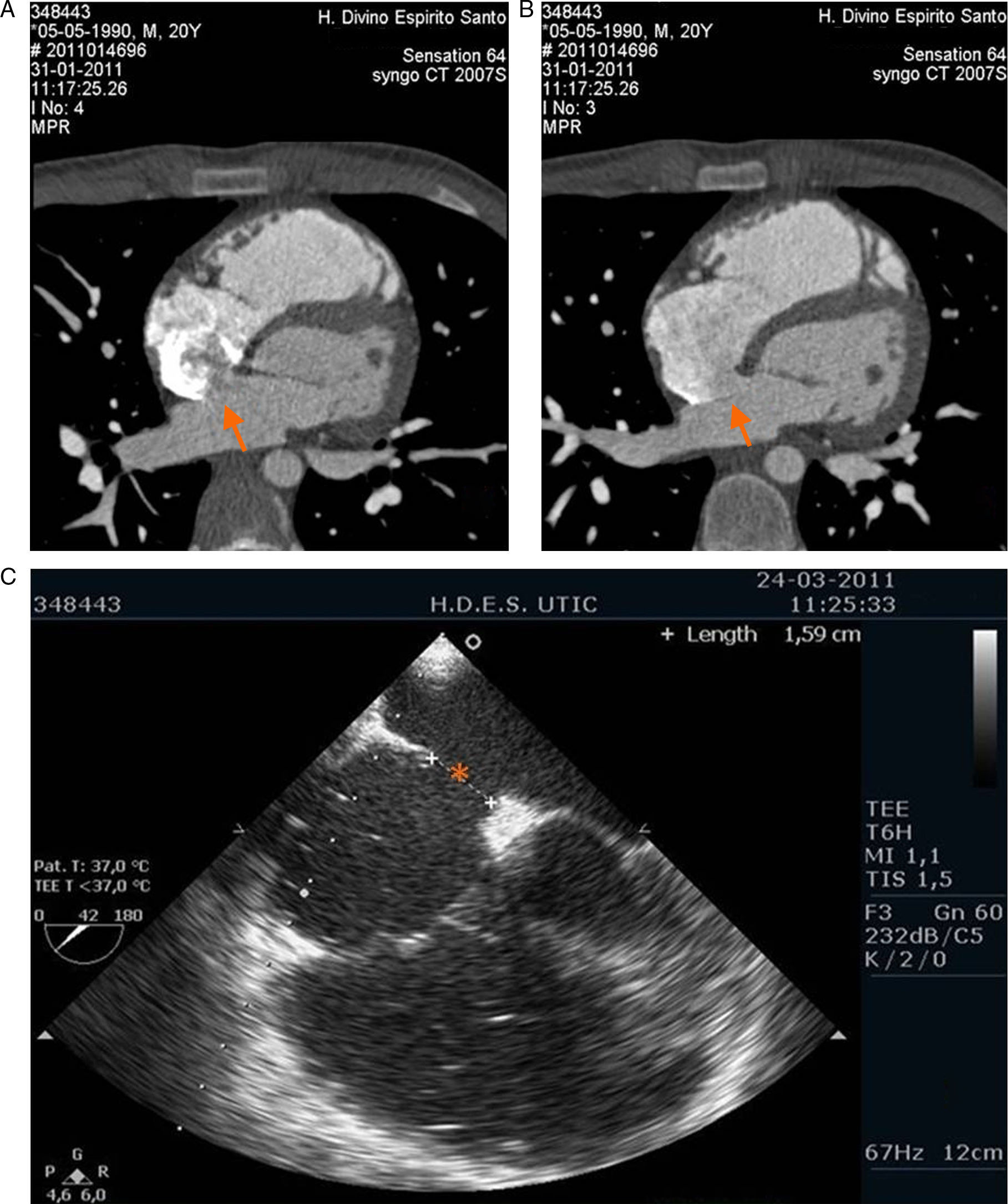
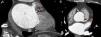
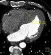
A 50-year-old man with a history of type 2 diabetes, permanent atrial fibrillation and chest pain, was referred for cardiac CT to exclude coronary disease. The exam revealed non-significant coronary disease, dilated LA and a subtraction image in the left atrial appendage suggestive of thrombus (Figure 2). The diagnosis could have been confirmed by a second scan a few minutes after the first or by transesophageal echocardiography, but these were not performed because the results would not have altered the therapeutic approach. The patient was prescribed oral anticoagulation.
Case 3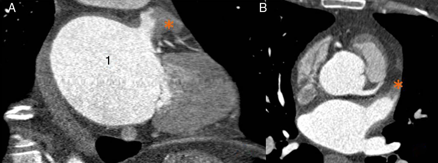
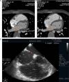
A 58-year-old man with known coronary disease, who had previously undergone angioplasty of the proximal anterior descending artery, experienced recurrence of angina and was accordingly referred for cardiac CT to assess stent patency. The exam excluded progression of coronary disease but revealed asymmetrical left ventricular hypertrophy and a ventricular septum of 36mm (Figure 3), compatible with hypertrophic cardiomyopathy. A cardioverter-defibrillator was implanted and to date no appropriate therapies have been recorded.
Discussion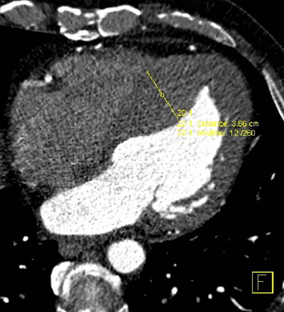
Cardiac CT is one of the most useful diagnostic techniques available to cardiologists. Advances in CT equipment, particularly in improved spatial resolution, shorter acquisition times and lower radiation doses, have made it possible to diagnose a growing range of cardiac disorders with increasing precision.
The authors present three cases in which cardiac CT was performed initially to exclude coronary disease but succeeded in diagnosing other cardiac disorders, leading to changes in treatment.
Several articles have been published on extracardiac findings diagnosed by cardiac CT.2 However, the intracardiac findings with prognostic impact presented here highlight the usefulness of this modality in diagnosing other cardiac disorders.
Conflicts of interestThe authors have no conflicts of interest to declare.
Please cite this article as: Almeida, C. Achados cardíacos com impacto prognóstico por AngioTC cardíaca. doi:10.1016/j.repc.2011.09.020.
