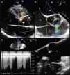
A 36-year-old man was referred to our hospital for a recent finding of a heart murmur. He had no relevant medical history except paroxysmal palpitations in the previous three months. Physical examination revealed a high-grade continuous rumbling murmur best heard at the left sternal border. Initial transthoracic echocardiography showed continuous flow between the aorta and the right ventricle (RV) (Figure 1A). The subsequent transesophageal echocardiogram (TEE) confirmed the presence of a tubular, windsock-like communication 4.5 mm in diameter between the right sinus of Valsalva (RSV) (displayed deformed) and the RV (Figure 1B and 1D, Video 1), with an effective regurgitant orifice of 0.11 cm2 in color flow Doppler (according to proximal isovelocity surface area measurement) and a systolic-diastolic flow in continuous wave Doppler (Figure 1C). There was also moderate aortic regurgitation (grade 3/4), with an eccentric jet directed toward the anterior mitral leaflet. Real-time three-dimensional (RT3D) TEE demonstrated the tubular morphology of the ruptured sinus (enlarged) and the direction of flow described above (Figure 2A and B, Video 2). Finally, multiplanar reconstruction color flow three-dimensional TEE showed a vena contracta area calculated by direct planimetry of 0.09 cm2 (Figure 2C). The patient underwent surgical defect closure with monofilament suture, leaving mild aortic regurgitation on intraoperative TEE.
(A) Transthoracic color Doppler echocardiography, parasternal long axis view, showing a continuous flow from the aorta to the right ventricle (arrow). (B) Transesophageal color Doppler echocardiography, mid-esophageal plane at 51°, showing the jet of the fistula (arrow) and moderate eccentric aortic regurgitation. (C) Continuous wave Doppler showing systolic-diastolic flow at the fistula level (arrow). (D) Transesophageal echocardiography, mid-esophageal plane at 124° (zoomed in on the aorta), showing a tubular communication between the right sinus of Valsalva and the right ventricle (arrow). Ao: aorta; LA: left atrium; LV: left ventricle; RA: right atrium; RV: right ventricle.
(A) Three-dimensional transesophageal echocardiography showing a tubular communication between the right sinus of Valsalva (enlarged) and the right ventricle. (B) Three-dimensional color flow showing the fistula (arrow) and the aortic regurgitation eccentric jet directed toward the anterior mitral leaflet. (C) Multiplanar reconstruction, three-dimensional color flow, showing a vena contracta area calculated by direct planimetry of 0.09 cm2. Ao: aorta; LV: left ventricle; RV: right ventricle.
A ruptured sinus of Valsalva communicating with the cardiac chambers is very uncommon, in our case probably secondary to congenital enlargement of the RSV. Supplementary information provided by RT3D echocardiography enabled appropriate assessment of the defect's anatomy.
Ethical disclosuresProtection of human and animal subjectsThe authors declare that no experiments were performed on humans or animals for this study.
Confidentiality of dataThe authors declare that they have followed the protocols of their work center on the publication of patient data and that all the patients included in the study received sufficient information and gave their written informed consent to participate in the study.
Right to privacy and informed consentThe authors declare that no patient data appear in this article.
Conflicts of interestThe authors have no conflicts of interest to declare.


