


Until a few years ago, technical limitations restricted the use of cardiac magnetic resonance (CMR) to anatomical assessment. The development of new imaging sequences and improvements in spatial and temporal resolution have widened indications for the technique.1 CMR is now a versatile imaging modality capable of assessing the anatomy, function, perfusion, flows and tissue characteristics of the heart and great vessels in a single exam that meets almost all the requirements of cardiac imaging. Images can be acquired non-invasively, reproducibly and relatively rapidly, unaffected by the acoustic window, and without exposing patients to ionizing radiation or iodinated contrast. These advances have led to a wider acceptance and steady growth in the use of the technique over the last 10 years.2 In Portugal, one of the obstacles to more frequent and appropriate use of CMR has been the lack of specific codes that accurately describe these examinations as they are currently performed. The coding of procedures, which is standard practice in modern medicine, is designed to aid statistical analyses, planning and evaluation of the needs of a given population, and the scheduling, invoicing and reimbursement of exams. Coding in most health care organizations entities in Portugal is outdated and overly general, with the exception of exams performed under the aegis of the National Health Service; these are coded and regulated by Order in Council no. 132/2009 and the codes referring to CMR are summarized in Table 1. This forms the basis for most of the recommendations in this document.
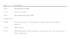
Codes for cardiac magnetic resonance exams performed under the aegis of the National Health Service, defined by Order in Council no. 132/2009.
| Code | Designation |
| 18111 | Morphological CMR |
| 18112 | Functional CMR |
| 18113 | Myocardial perfusion CMR |
| Supplements | |
| 18240 | Post-processing (e.g. 3D sequence, volume rendering, functional analysis) |
| 18210 | MRI contrast |
| 18234 | MR angiography with gadolinium, any location (this code excludes any other supplement) |
CMR: cardiac magnetic resonance; MR: magnetic resonance.
It should be borne in mind that the term “cardiac magnetic resonance” covers a large number of complementary but different techniques. The attending physician is responsible for tailoring the protocol of each exam according to clinical suspicion and the particular characteristics of the patient, choosing the appropriate techniques, sequences and views for each situation. The result is considerable diversity in the parameters assessed, the sequences used and exam duration, which is difficult to frame within a single code.
The aim of this document, which has been drawn up by physicians experienced in the use of CMR, is to make recommendations for updating and standardizing the codes for this imaging modality in Portugal. Guidance on which techniques and codes should be used in the most common clinical scenarios is also provided.
Coding of cardiac magnetic resonance examsThe proposed codes are summarized in Table 2. A brief description of CMR techniques and their clinical uses follows.
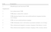
New codes proposed for cardiac magnetic resonance.
| Code | Designation |
| 1 | Morphological and functional CMR |
| Supplements | |
| 2 | Late enhancement CMR |
| 3 | CMR phase contrast flow analysis |
| 4 | CMR pharmacological stress myocardial perfusion imaging (includes cost of drug) |
| 5 | CMR rest myocardial perfusion imaging |
| 6 | CMR dobutamine stress wall motion assessment (includes cost of drug) |
| 7 | Three-dimensional magnetic resonance imaging of the thoracic vessels |
| 8 | Three-dimensional magnetic resonance imaging of the coronary arteries |
All supplementary codes except 3, 6 and 8 assume the administration of paramagnetic contrast.
This, the basic code, covers the morphological and functional assessment that is essential in any CRM exam. It should use dynamic images from balanced steady-state free precession (bSSFP) or gradient-recalled echo sequences and static images from turbo spin-echo or short-tau inversion recovery sequences. The information obtained includes quantification of ventricular volumes and function, and quantification of left ventricular mass, assessment of wall motion and tissue characterization, all without the need for contrast agents, which is useful in the differential diagnosis of a variety of heart diseases.
Late enhancement studiesOne of the main advantages of CRM is non-invasive detection of the presence, extent and pattern of myocardial fibrosis, based on T1-weighted inversion recovery sequences with image acquisition around 10 minutes after the administration of paramagnetic contrast. Late enhancement study is indicated in the investigation of cardiomyopathy and myocarditis, ischemic heart disease (to assess myocardial viability and to detect subclinical infarction), cardiac masses, pericardial disease, and other conditions. The many clinical applications of this technique mean that it is used in most CMR exams.
Phase contrast flow analysisBlood flow can be assessed by CRM using phase contrast methods, which is particularly useful in the investigation of valve disease and congenital heart defects. Quantitative flow analysis requires special post-processing software.
Pharmacological stress myocardial perfusion imagingMyocardial perfusion imaging is usually based on assessment of the dynamics of the first pass of a bolus of paramagnetic contrast using fast T1-weighted sequences. Stress in this context refers to the administration of a coronary vasodilator, normally adenosine or dipyridamole, to induce maximum hyperemia. The presence of obstructive atherosclerotic lesions reduces the vasodilator response, leading to a regional asymmetry in myocardial perfusion. Diagnosis of reduced coronary flow reserve is based on comparison of perfusion in different vascular territories and of images obtained during stress and at rest. In some countries, assessment of myocardial perfusion under pharmacological stress is now one of the main reasons for referral for CMR.3 The indications for this technique are similar to those for myocardial perfusion scintigraphy (MPS), but CMR has better spatial resolution and does not involve ionizing radiation. In a recent study, CMR was superior to MPS in the diagnosis of coronary heart disease4; the use of CMR to detect ischemic disease is expected to increase considerably in the coming years.
Rest myocardial perfusion imagingRest myocardial perfusion imaging is used for comparisons with pharmacological stress myocardial perfusion imaging to assess the reversibility of perfusion defects and in the investigation of cardiac masses to determine the extent of vascularization. The same sequences are used as in stress imaging.
Dobutamine stress wall motion imagingThis technique assesses dobutamine-induced wall motion abnormalities in a similar way to stress echocardiography. Dobutamine is administered via a peripheral vein in increasing doses up to a maximum of 40 μg/kg/min in four or five stages each of 3-5 min, and can be supplemented by atropine to attain the target heart rate. Wall motion is assessed at each stage using bSSFP cine sequences. The use of dobutamine requires careful monitoring of blood pressure, cardiac rhythm and symptoms; the attending physician must be prepared to deal with any complications, particularly arrhythmias or symptoms of myocardial ischemia. Low-dose dobutamine CMR (up to 10 μg/kg/min) is an alternative to late enhancement CMR to assess myocardial viability and the integrity of the myocardial contractile apparatus, or when the transmurality of one or more segments on late enhancement is intermediate or unclear. Dobutamine stress CMR is logistically complex, time-consuming and not without risk, and is therefore now used less to assess ischemia and myocardial viability than vasodilator stress imaging and late enhancement studies, respectively. It is generally used only in cases in which paramagnetic contrast is contraindicated. Despite these limitations, dobutamine stress CMR has higher diagnostic accuracy than dobutamine stress echocardiography,5 particularly when there is a poor acoustic window.
Three-dimensional magnetic resonance imaging of the thoracic vesselsAngiography of the great vessels is usually performed following administration of a bolus of paramagnetic contrast, acquisition time being set to coincide with the passage of contrast through the area of interest. The technique provides accurate three-dimensional angiographic images of the aorta, pulmonary arteries and their branches, pulmonary veins, and other vessels.
Three-dimensional magnetic resonance imaging of the coronary arteriesThis technique is little used in clinical practice, as limitations of spatial and temporal resolution mean that only the proximal segments of the coronary arteries can be assessed. Its main application is to exclude anomalous coronary artery origin and to assess coronary aneurysms in Kawasaki disease; it is not usually indicated in the study of atherosclerotic coronary disease.
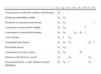
Association of codes and components of cardiac magnetic resonance imagingThe complexity of CMR and the fact that most physicians are unfamiliar with the technique mean that it is generally impractical for the physician requesting the exam to specify beforehand which components are required to clarify the particular clinical questions to be answered. Given this situation, we propose a list of components and codes for the most common clinical indications (Table 3), in a similar fashion to the international guidelines.6
Components of cardiac magnetic resonance imaging and the corresponding codes to include for the most common clinical indications.
| 1 | 2 | 3 | 4 | 5 | 6 | 7 | 8 | |
| Assessment of ventricular volumes and function | ¿ | |||||||
| Cardiomyopathy/Myocarditis | ¿ | ¿ | ||||||
| Exclusion of structural heart disease | ¿ | ¿ | a | |||||
| Assessment of myocardial viability | ¿ | ¿ | b | |||||
| Assessment of myocardial ischemia | ¿ | ¿ | ¿ | ¿ | c | |||
| Valve disease | ¿ | a | ¿ | |||||
| Congenital heart defects | ¿ | ¿ | ¿ | ¿ | a | |||
| Pericardial disease | ¿ | ¿ | ||||||
| Assessment of cardiac masses | ¿ | ¿ | ¿ | |||||
| Diseases of the thoracic vessels | ¿ | ¿ | ¿ | |||||
| Assessment before or after ablation of atrial fibrillation | ¿ | ¿ | a | ¿ |
1: morphological and functional CMR; 2: late enhancement CMR; 3: CMR phase contrast flow analysis; 4: CMR pharmacological stress myocardial perfusion imaging; 5: CMR rest myocardial perfusion imaging; 6: CMR dobutamine stress wall motion assessment; 7: three-dimensional magnetic resonance imaging of the thoracic vessels with gadolinium; 8: three-dimensional magnetic resonance imaging of the coronary arteries.
The costing of CMR exams is beyond the scope of this document and will be subject to negotiation between funding bodies and providers. However, certain guiding principles should be borne in mind in order to ensure that CRM exams are simultaneously affordable for funders and financially viable for service providers. The authors suggest that the figures set out in Order in Council no. 132/2009 should be taken as a reference, and that any adjustments should take the following factors into account:
- 1
The duration and complexity of the exam compared to other magnetic resonance (MR) exams; for example, a CMR study usually takes 3-4 times longer than MR study of the knee or skull. A comparison of the duration and complexity of these exams can be found in the joint recommendations of the British Society of Cardiovascular Imaging and the British Society of Cardiovascular Magnetic Resonance on codes and tariffs for CMR (available at http://www.bsci.org.uk/ct-cmr-tariffs);
- 2
The need for specific acquisition sequences. State-of-the-art CMR exams involve sequences that entail software license costs for the centers that provide this service;
- 3
The need for post-processing, particularly for determination of ventricular volumes and function, and the specialized software required. This should be performed by a physician;
- 4
Comparison between CMR and other non-invasive imaging modalities including transesophageal echocardiography, myocardial perfusion scintigraphy and cardiac computed tomography angiography. For example, in our experience a CMR exam to assess myocardial ischemia will cost more than cardiac computed tomography angiography but little more than stress-rest myocardial perfusion scintigraphy.
In this consensus document, recommendations are made for updating and standardizing CMR codes in Portugal. Guidance on which techniques and codes should be used in the most common clinical scenarios is also provided.
Ethical disclosuresProtection of human and animal subjectsThe authors declare that no experiments were performed on humans or animals for this study.
Confidentiality of dataThe authors declare that no patient data appear in this article.
Right to privacy and informed consentThe authors have obtained the written informed consent of the patients or subjects mentioned in the article. The corresponding author is in possession of this document.
Conflicts of interestThe authors have no conflicts of interest to declare.
Please cite this article as: Ferreira AM, et al. Documento de Consenso sobre Codificação de exames de Ressonância Magnética Cardíaca em Portugal. Rev Port Cardiol. 2012. doi:10.1016/j.repc.2012.08.003.
