Journal Information
Vol. 40. Issue 11.
Pages 891-893 (November 2021)
Share
Share
Download PDF
More article options
Visits
2653
Vol. 40. Issue 11.
Pages 891-893 (November 2021)
Snapshot
Open Access
Cardiac resynchronization system implantation guided by three-dimensional electroanatomic mapping
Implantação de sistema de ressincronização cardíaca guiada por mapeamento eletroanatómico 3D
Visits
2653
Pedro Silva Cunha
Corresponding author
psilvacunha@sapo.pt

Corresponding author.
, Hélder Santos, Mário Martins Oliveira
Arrhythmology, Pacing and Electrophysiology Unit, Cardiology Service, Santa Marta Hospital, Central Lisbon Hospital University Center (CHULC), Portugal
This item has received

Under a Creative Commons license
Article information
Full Text
Bibliography
Download PDF
Statistics
Figures (4)
Show moreShow less
Full Text
Clinical case and implantation procedure

A 44-year-old man with a diagnosis of non-ischemic dilated cardiomyopathy was referred for implantation of a cardiac resynchronization therapy (CRT) system with cardioverter-defibrillator (CRT-D) as primary prevention. The procedure was performed using the EnSite™ Precision™ (Abbott).

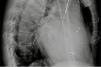
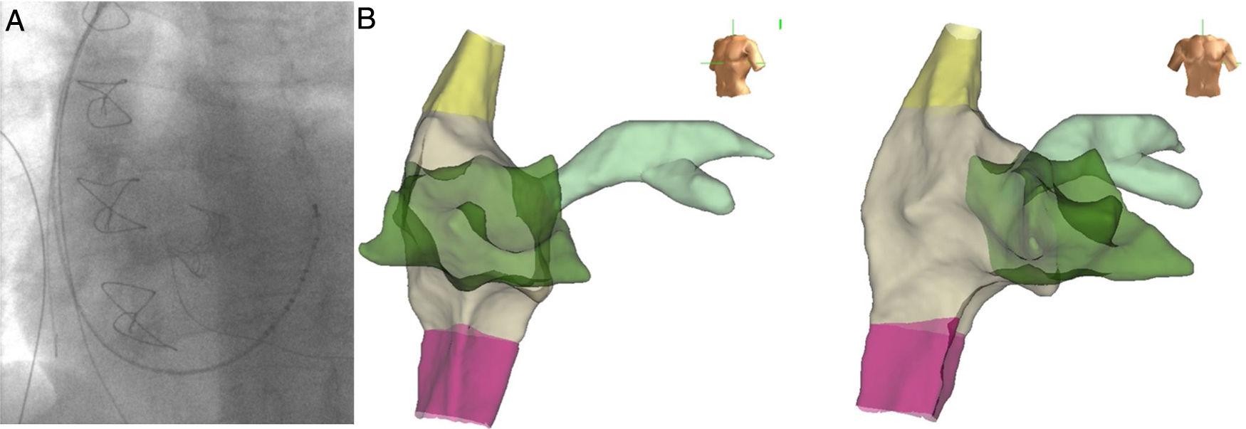
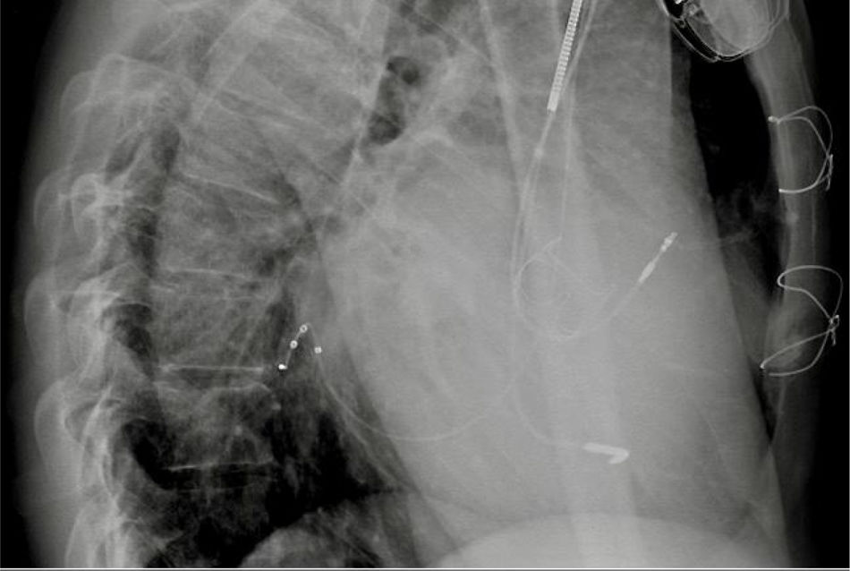
Via a subclavian vein approach, a 5F decapolar Response™ CSL electrophysiology catheter (Abbott) was used to create a three-dimensional (3D) model.1 After obtaining an endocardial shell the catheter was used to cannulate the coronary sinus (CS) (Figure 1). Under guidance by the electroanatomic system and very low dose fluoroscopy (to confirm that there was no catheter entanglement), a bipolar right ventricular (RV) lead was positioned in the RV apical septum. To continuously monitor the lead position (and identify possible lead dislodgment), the tip and ring poles were left connected to the EnSite system. The CS catheter was withdrawn and a sheath was used to catheterize the CS with the aid of the anatomic location on the 3D system. A guidewire (VisionWire; Biotronik) was inserted through the delivery system and connected to the EnSite system, and was used to map the anatomy and electrical activation of the CS branches (Figure 2). A branch was selected and the LV lead was placed in the latest site of ventricular activation (Figures 3 and 4). Using guidance by the navigation system and minimal fluoroscopy (hybrid approach), the right atrial lead was placed in a right appendage position, with real-time location of the other catheters (to check for possible displacement during manipulation of the atrial lead). Total procedure time was 96 min.

Figure 1.

(A) Fluoroscopy image of the decapolar Response™ CSL electrophysiology catheter inside the coronary sinus; (B) simultaneous EnSite three-dimensional electroanatomic reconstruction of the anatomy of the right heart chambers and the coronary sinus, without fluoroscopy.

(0.09MB).
Figure 2.

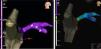
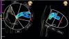
(A) Real-time visualization of catheter location (red arrow pointing to right ventricular lead, and yellow arrow to right atrial lead) and the VisionWire (Biotronik) inside the coronary sinus branches (white arrows); (B): electrical activation mapping of coronary sinus branches with guidewire and identification of the latest ventricular activation (mid segment of the lateral branch, dark blue).

(0.17MB).
Figure 3.

Coronary sinus activation mapping and final position of the left ventricular lead (red arrow).

(0.3MB).
Figure 4.

Chest X-ray (left lateral view) with lead position 24 hours after implantation.

(0.06MB).

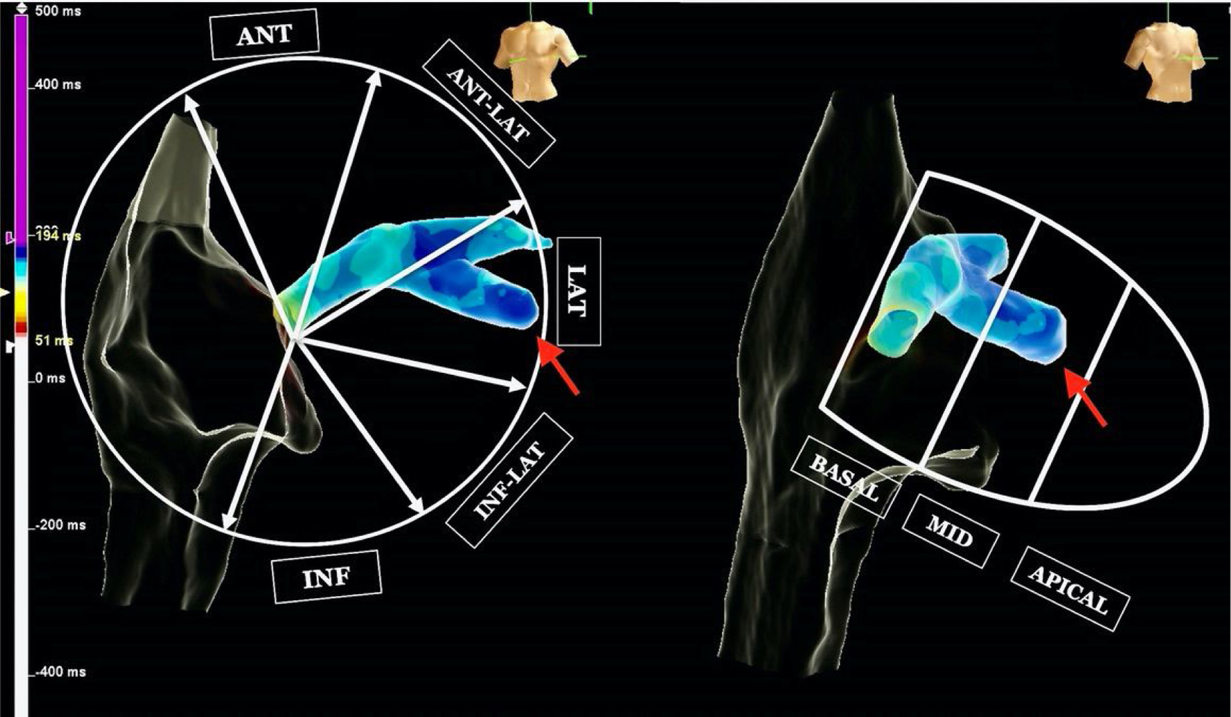
In this case the use of a 3D electroanatomic system with the VisionWire additionally enabled LV activation to be mapped, and guided optimal LV lead implantation toward segments of delayed LV activation (QRS width reduced from 165 ms to 100 ms with CRT on). This implantation approach allows a greater reduction in fluoroscopy and angiography times2–4 compared with the traditional technique of CRT implantation.

Funding

This work did not receive any specific grant from funding agencies in the public, commercial, or not-for-profit sectors.

Conflicts of interest

The authors have no conflicts of interest to declare.

References
[1]
C. Eitel, G. Hindricks, N. Dagres, et al.
EnSite velocity cardiac mapping system: a new platform for 3D mapping of cardiac arrhythmias.
Expert Rev Med Dev, 7 (2010), pp. 185-192
[2]
M. Giaccardi, A. Del Rosso, V. Guarnaccia, et al.
Near-zero X-ray in arrhythmia ablation using a 3-dimensional electroanatomic mapping system: a multicenter experience.
Heart Rhythm, 13 (2016), pp. 150-156
[3]
A. Mina, Warnecke N:.
Near zero fluoroscopic implantation of BIV ICD using electro-anatomical mapping.
Pacing Clin Electrophysiol, 36 (2013), pp. 1409-1416
[4]
A. Colella, M. Giaccardi, T. Colella, et al.
Zero x-ray cardiac resynchronization therapy device implantation guided by a nonfluoroscopic mapping system: a pilot study.
Heart Rhythm, 13 (2016), pp. 1481-1488
Copyright © 2021. Sociedade Portuguesa de Cardiologia
Idiomas
Revista Portuguesa de Cardiologia (English edition)
Article options
Tools
en pt

Are you a health professional able to prescribe or dispense drugs?

Você é um profissional de saúde habilitado a prescrever ou dispensar medicamentos

By checking that you are a health professional, you are stating that you are aware and accept that the Portuguese Journal of Cardiology (RPC) is the Data Controller that processes the personal information of users of its website, with its registered office at Campo Grande, n.º 28, 13.º, 1700-093 Lisbon, telephone 217 970 685 and 217 817 630, fax 217 931 095, and email revista@spc.pt. I declare for all purposes that the information provided herein is accurate and correct.