The authors present the case of a 39-year-old man with a history of unspecified congenital heart disease, who was asymptomatic and had never undergone any intervention or cardiological follow-up.
He was admitted for insidious weight loss, fatigue and fever. He presented a IV/VI systolic murmur over the precordium, hepatosplenomegaly and lower limb edema. Streptococcus viridans was isolated from blood cultures and multiple foci of pulmonary and splenic emboli were identified.
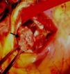
Transthoracic and transesophageal echocardiography revealed a restrictive subpulmonary ventricular septal defect (VSD) and infundibular pulmonary stenosis (Figure 1 and Video 1), with multiple vegetations related to the VSD involving the right coronary cusp of the aortic valve and protruding into the right ventricular outflow tract (RVOT) (Figure 2 and Videos 2 and 3). Vegetations were also observed adhering to the pulmonary valve and the pulmonary artery wall, probably due to jet lesion (Figure 3 and Video 4). A diagnosis of infectious endocarditis was made and the patient underwent surgery for removal of the vegetations (Figure 4), VSD closure, enlargement of the RVOT and pulmonary valvuloplasty without use of prosthetic material. His postoperative course was good, with no significant residual lesions, and there were no signs of heart failure or recurrence of endocarditis after two years of follow-up.
This case is an example of congenital heart disease with multiple defects with an indolent course that was manifested by aggressive infectious endocarditis involving the left and right valves and with high embolic risk. It also highlights the success of surgical intervention to simultaneously remove the infected material and repair the congenital defects.
Ethical disclosuresProtection of human and animal subjectsThe authors declare that the procedures followed were in accordance with the regulations of the relevant clinical research ethics committee and with those of the Code of Ethics of the World Medical Association (Declaration of Helsinki).
Confidentiality of dataThe authors declare that they have followed the protocols of their work center on the publication of patient data.
Right to privacy and informed consentThe authors have obtained the written informed consent of the patients or subjects mentioned in the article. The corresponding author is in possession of this document.
Conflicts of interestThe authors have no conflicts of interest to declare.
Please cite this article as: Faustino M, Freitas A, Oliveira Soares A, Fragata J, Gil VM, Morais C. Cardiopatia congénita do adulto, substrato para endocardite infeciosa. Rev Port Cardiol. 2015;34:145–146.


