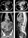
A 62-year-old woman undergoing control exams following resection of a left renal tumor two years previously reported only dyspnea on moderate to strenuous exertion. Computed tomography (CT) angiography of the chest, abdomen and pelvis revealed an idiopathic aneurysmal dilatation of the proximal inferior vena cava (IVC) next to the right atrium, 56 mm on its largest axis (type I in the Gradman and Steinberg classification) (Figure 1). The middle, right and left hepatic veins drained into the aneurysmatic area, which was herniated into the thoracic region with part of the hepatic segment. Distal to the aneurysm, in the intrahepatic portion, the IVC was practically collapsed, with a very small caliber, for about 35 mm. In the subhepatic region, into which the other abdominal and pelvic veins drained, the IVC had a normal appearance and caliber.
Computed tomography angiography of the chest, abdomen and pelvis, without contrast (A) and with contrast in the venous phase (B-D), showing aneurysmal dilatation of the proximal inferior vena cava next to its opening into the right atrium, reaching 56 mm in its largest diameter (A and B, straight white arrow). The middle, right and left hepatic veins drained into the aneurysmatic area, which was herniated into the thoracic region with part of the hepatic segment (C, white circle). Distal to the aneurysm, in the intrahepatic portion, the IVC was practically collapsed, with a very small caliber, for about 35 mm (D, black arrow). In the subhepatic region, into which the other abdominal and pelvic veins drained, the IVC had a normal appearance and caliber (C, curved white arrow).
Venous aneurysm, especially of the IVC, is very rare, and its etiology, risk factors and prognosis are poorly understood. Inflammatory disease, thrombosis, trauma and long-standing right heart failure are possible risk factors. It may also be the result of embryonic malformation. Clinical presentation is variable and it may be a chance finding in asymptomatic patients.
CT, magnetic resonance angiography and venography provide detailed images of the aneurysmal structure. Surgery is indicated for all such patients if symptomatic, but an incidental diagnosis in an asymptomatic patient is a challenging situation.
Ethical disclosuresProtection of human and animal subjectsThe authors declare that no experiments were performed on humans or animals for this study.
Confidentiality of dataThe authors declare that no patient data appear in this article.
Right to privacy and informed consentThe authors declare that no patient data appear in this article.
Conflicts of interestThe authors have no conflicts of interest to declare.
Please cite this article as: Luís Duarte M, Abreu BB, Silva AQ, Prado JL, Silva MQ. Aneurisma idiopático da veia cava inferior – diagnóstico tomográfico. Rev Port Cardiol. 2017;36:781–782.


