


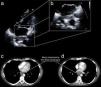
In this Image in Cardiology we describe the case of a lymphoma of the esophagus discovered by transthoracic echocardiography.
A 50-year-old Caucasian male was referred to our echocardiography laboratory complaining of epigastric pain and exertional dyspnea. Standard transthoracic echocardiography showed an ovoid, heterogeneous mass measuring about 4 cm×2 cm extending between the left atrium and the descending aorta (Figure 1a and b, Movie clip 1), not causing compression of the left atrium or the pulmonary veins.
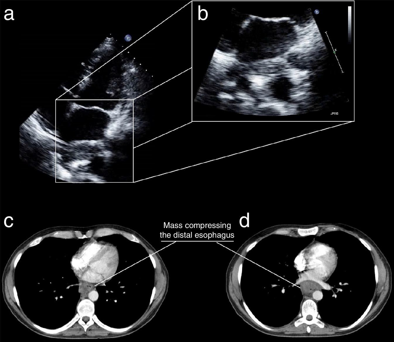
Total body computed tomography with iodinated contrast was promptly performed, which showed a hypodense lesion, measuring about 7 cm, of a thickened (about 3 cm) distal esophagus (Figure 1c and d), accompanied by enlarged satellite lymph nodes.
Significant narrowing of the distal esophagus was also revealed by Gastrografin swallow.
Endoscopic examination was performed showing an ulcerated mass with circumferential growth constricting the distal third of the esophageal lumen. Multiple biopsies were obtained and histologic examination showed large, atypical lymphoid cells positive for CD30 and ALK.
The mass was accordingly diagnosed as a primary anaplastic large cell lymphoma of the esophagus.
Ethical disclosuresProtection of human and animal subjectsThe authors declare that no experiments were performed on humans or animals for this study.
Confidentiality of dataThe authors declare that no patient data appear in this article.
Right to privacy and informed consentThe authors declare that no patient data appear in this article.
Conflicts of interestThe authors have no conflicts of interest to declare.
We are grateful to sonographer Chiadon Secka, who performed the transthoracic echocardiography.
