The CAD-RADSTM classification was recently introduced in an attempt to standardize coronary computed tomography angiography (CCTA) reports and to provide recommendations for further management. The aim of this study was to assess how additional cardiac investigations were being ordered before the introduction of the CAD-RADS classification in a tertiary hospital's CCTA reports.
MethodsWe conducted a single-center retrospective analysis of 200 patients (103 women, mean age 59±13 years) who underwent CCTA for suspected or known coronary artery disease prior to the systematic introduction of the CAD-RADS classification in the reports. For each case, we assessed whether further cardiac investigation was requested after CCTA and what type of test was performed (functional testing, invasive coronary angiography or viability testing).
ResultsThe majority of patients (n=158; 79%) were classified as CAD-RADS 0-2. In patients with lower (0-2) or higher (4 or 5) scores, further testing was in accordance with CAD-RADS recommendations in 98% of cases (n=168). In patients with CAD-RADS 3 (intermediate stenosis), functional testing was requested as recommended in only 36% of cases (n=5), while 50% (n=7) proceeded directly to invasive coronary angiography. In patients in whom CCTA was non-diagnostic, most did not undergo further cardiac investigation.
ConclusionIn patients with CAD-RADS classifications at the ends of the spectrum, additional cardiac investigation after CCTA was almost always in accordance with the recommendations. However, in patients with intermediate scores, invasive coronary angiography prevailed over functional testing.
A classificação CAD-RADS® foi recentemente introduzida com o objetivo de padronizar os relatórios da AngioTC coronária e fornecer recomendações sobre a investigação futura. O objetivo deste trabalho foi avaliar qual o tipo de investigação cardíaca adicional solicitada antes da introdução da classificação CAD-RADS® nos relatórios de AngioTC coronária de um hospital terciário.
MétodosAnálise retrospetiva de centro único em que foram avaliados 200 doentes (103 mulheres, idade média 59±13 anos) que realizaram AngioTC por suspeita ou avaliação de doença coronária, antes da introdução sistemática da classificação CAD-RADS® nos relatórios. Em cada caso avaliou-se se foi realizada investigação cardíaca adicional após a AngioTC e qual o tipo de exame requisitado (avaliação funcional de isquemia, angiografia coronária invasiva ou teste de viabilidade).
ResultadosA maioria dos doentes (n = 158; 79%) foram classificados como CAD-RADS® 0 - 2. Nos doentes com pontuação mais baixa (0-1) ou mais elevada (4-5), a investigação realizada foi concordante com as recomendações em 98% dos casos (n = 168). Em doentes com CAD-RADS® = 3 (estenose intermédia), os testes funcionais foram requisitados em apenas 36% dos casos (n = 5), enquanto 50% (n = 7) realizaram diretamente angiografia coronária invasiva. Nos doentes em que o estudo não foi diagnóstico, a maioria não realizou nova investigação cardíaca.
ConclusãoNos doentes com classificação CAD-RADS® nos extremos da pontuação, a avaliação cardíaca adicional após realização de AngioTC coronária foi muito semelhante àquela prevista pelo score. Contudo, nos doentes com classificação intermédia, a coronariografia prevaleceu sobre o teste funcional.
The increasing availability and broadening indications for coronary computed tomography angiography (CCTA) have highlighted the need to establish a common language in reporting and to provide guidance for further clinical management. In this context, three medical societies recently issued a consensus document introducing the Coronary Artery Disease - Reporting and Data System (CAD-RADSTM) classification, aimed at facilitating communication of test results to referring physicians using simple terminology and offering guidance for subsequent patient management.1,2 The impact of the systematic introduction of the CAD-RADS classification in CCTA reports remains to be established, but will ultimately depend on its acceptance and how far real-world post-test management is from proposed care. The aim of this study was to assess how additional cardiac investigations were being ordered before the introduction of the CAD-RADS classification in a tertiary hospital's CCTA reports.
MethodsStudy populationWe conducted this retrospective study in a tertiary cardiovascular center, where consecutive patients undergoing CCTA for suspected or known coronary artery disease (CAD) were included in an observational registry from October 2015 to September 2016. We excluded patients with recent (<6 months) invasive coronary angiography who underwent CCTA to assess bridging segments or bypass grafts that could not be successfully catheterized (n=6). The final study population consisted of 200 individuals. All patients gave written informed consent.
Data on patient demographics, symptoms and medical history were obtained from a structured pre-test interview, supplemented with information provided by the referring physician and electronic medical records. CCTA was ordered by staff cardiologists or cardiac surgeons and was performed prior to the systematic introduction of the CAD-RADS classification in the reports. For each case, we assessed whether further cardiac investigation was requested after CCTA and what type of test was performed: functional testing, invasive coronary angiography (ICA) or viability testing. Functional testing included exercise electrocardiography, stress echocardiography, stress single-photon emission computed tomography (SPECT), stress cardiac magnetic resonance (CMR), and stress positron emission tomography (PET). Viability testing included low-dose dobutamine echocardiography, rest SPECT, CMR or PET. Patients were followed until the referring physician made the decision to order or forgo further testing. Each patient's pre-test probability of obstructive CAD was calculated according to European Society of Cardiology guidelines.3
Coronary computed tomography angiography and image analysisCCTA was performed on a dual-source 64-slice computed tomography scanner (Somatom Definition®, Siemens Healthineers, Germany) in accordance with the Society of Cardiovascular Computed Tomography guidelines.4 Unless contraindicated, patients received a single dose of 0.5mg sublingual nitroglycerin, and oral or intravenous beta-blocker if heart rate was >70 beats per minute. A bolus of 80-100ml intravenous contrast agent, followed by saline solution, was injected through an arm vein at a flow rate of 5ml/s.
Images were reconstructed with electrocardiographic gating to obtain optimal, motion-free image quality. Radiation dose reduction strategies were employed when feasible. Effective radiation dose for CCTA was determined using the dose-length product with an organ-specific conversion factor of 0.014 mSv/mGy.cm.5 Total calcium score was calculated with dedicated software and expressed as Agatston score.6 Calcium scoring was not performed in patients with coronary stents.
A cardiologist and/or radiologist with more than five years’ experience in CCTA analyzed all scans on an Aquarius workstation (Terarecon Inc., Foster City, CA, USA) using axial images, multiplanar reconstructions, and maximum intensity projections, as appropriate. Coronary stenosis severity was assessed by visual estimation.
CAD-RADS classificationThe CAD-RADS classification system was applied on a per-patient basis representing the highest-grade coronary artery stenosis documented by CCTA. A summary of the CAD-RADS classification is presented in Table 1.
CAD-RADS classification.
| Classification | Definition | Further investigation |
|---|---|---|
| 0 | Absence of CAD (no plaque or stenosis) | None |
| 1 | Minimal stenosis or plaque without stenosis (1-24%) | None |
| 2 | Mild stenosis (25-49%) | None |
| 3 | Moderate stenosis (50-69%) | Consider functional assessment |
| 4 | 4A: 70-99% stenosis4B: LM >50% or 3-vessel obstructive (≥70%) disease | 4A: Consider functional assessment or ICA4B: ICA is recommended |
| 5 | Total occlusion (100%) | Consider ICA and/or viability assessment |
| Na | Non-diagnostic study | Additional or alternative assessment may be needed |
Study is not fully assessable or is non-diagnostic.
CAD-RADS does not apply to smaller vessels (<1.5mm in diameter).
Modifiers: N (non-diagnostic), S (stent), G (graft), V (vulnerability).
CAD: coronary artery disease; ICA: invasive coronary angiography; LM: left main.
Adapted from Cury et al.1
Continuous variables are expressed as mean ± standard deviation (SD), or median and interquartile range for variables with non-normal distributions. Normality of distribution was assessed with the Kolmogorov-Smirnov test. Categorical variables are presented as percentage. Two-sided p values <0.05 were considered statistically significant. The statistical analysis was performed with IBM SPSS version 22.0 for Mac OS X.
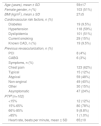
ResultsBaseline patient characteristicsThe clinical characteristics of the study population are summarized in Table 2. The main reasons for undergoing CCTA were suspected CAD in symptomatic patients with low-to-intermediate pre-test probability (74%), preoperative exclusion of CAD (11%), and left ventricular systolic dysfunction of unknown etiology (7.5%). A total of 19 patients had previously documented CAD and 12 had undergone myocardial revascularization. In roughly one third of cases (n=70), CCTA followed some form of ischemia testing (60 exercise electrocardiogram, eight SPECT and two stress echocardiography).
Clinical characteristics of the study population.
| Age (years), mean ± SD | 59±17 |
| Female gender, n (%) | 103 (51%) |
| BMI (kg/m2), mean ± SD | 27±5 |
| Cardiovascular risk factors, n (%) | |
| Diabetes | 19 (9.5%) |
| Hypertension | 118 (59%) |
| Dyslipidemia | 101 (51%) |
| Current smoking | 29 (15%) |
| Known CAD, n (%) | 19 (9.5%) |
| Previous revascularization, n (%) | |
| PCI | 8 (4%) |
| CABG | 6 (3%) |
| Symptoms, n (%) | |
| Chest pain | 123 (62%) |
| Typical | 15 (12%) |
| Atypical | 59 (48%) |
| Non-anginal | 49 (40%) |
| Other | 30 (15%) |
| Asymptomatic | 47 (24%) |
| PTPa(n=102) | |
| <15% | 12 (12%) |
| 15%-65% | 80 (78%) |
| 66%-85% | 9 (8.8%) |
| >85% | 1 (1.0%) |
| Heart rate, beats per minute, mean ± SD | 65±10 |
CCTA characteristics are depicted in Table 3. Obstructive CAD (≥50%) was detected in 28 patients (14%). Among these, the left anterior descending artery was involved in 54%, the right coronary artery in 43% and the circumflex artery in 32%.
Coronary computed tomography angiography characteristics of the study population.
| Prospective gating | 97 (49%) |
| Effective radiation dose (mSv), median (IQR) | 4.3 (2.9, 6.8) |
| Agatston CAC score, median (IQR) | 1.9 (0, 74) |
| Coronary circulation dominance, n (%) | |
| Right | 165 (83%) |
| Left | 18 (9%) |
| Codominance | 17 (9%) |
| Number of obstructive lesions (≥50%), n (%) | 28 (14%) |
| 1 | 17 (61%) |
| 2 | 8 (29%) |
| 3 | 3 (11%) |
| CAD-RADS classification, n (%) | |
| 0 – No plaque or stenosis | 77 (39%) |
| 1 – Minimal stenosis | 38 (19%) |
| 2 – Mild stenosis | 43 (22%) |
| 3 – Moderate stenosis | 14 (7%) |
| 4A – Severe stenosis | 9 (4.5%) |
| 4B – LM >50% or 3-vessel disease ≥70% | 1 (0.5%) |
| 5 – Total occlusion | 4 (2%) |
| N – Non-diagnostic study | 14 (7%) |
CAC: coronary artery calcium; IQR: interquartile range; LM: left main.
The distribution of patients according to the CAD-RADS classification and additional cardiac investigations is shown in Figure 1. Most patients (79%) were classified with the lowest scores (score 0-2). In patients with low (0-2) or high (4 or 5) CAD-RADS scores, further investigation was in accordance with that suggested by the classification in 98% of cases (n=168). Only four patients with CAD-RADS 2 underwent ICA (n=2) or functional testing (n=2).
In patients with intermediate grade lesions (CAD-RADS 3), functional testing was requested as recommended in only 36% of cases (n=5), with 50% (n=7) undergoing ICA and the remainder no further investigation (14%; n=2). In four of the seven patients with CAD-RADS 3 who underwent ICA, fractional flow reserve (FFR) was measured during the procedure. In patients in whom CCTA was non-diagnostic (no apparent obstructive CAD but with non-assessable segments), most (64%; n=9) did not undergo further cardiac investigation.
A subgroup analysis was carried out excluding patients with previous known CAD (n=19). As in the main analysis, coronary angiography remained the most requested exam in patients with an intermediate score (54%; n=7), and functional assessment was the second most frequently requested (38%; n=5). Additional cardiac studies requested were also similar for other CAD-RADS scores(Figure 2A).
In patients with previous known CAD (n=19), the main reasons for requesting CCTA were (1) for assessment of previously documented non-significant CAD in asymptomatic patients, patients with positive exercise test or with atypical symptoms (n=12); (2) assessment of grafts (n=5); and (3) assessment of large-caliber proximal stents (n=2). In this population, 42% of patients had the lowest scores (0-2), 26% the highest scores (4 and 5) and 26% (n=5) had non-diagnostic studies, of whom two patients underwent ICA. Among patients with CAD-RADS 4 or 5, one patient underwent functional testing and the remainder underwent ICA (Figure 2B).
DiscussionThe aim of this study was to assess how additional cardiac investigations were being ordered before the introduction of the CAD-RADS classification in CCTA reports in a tertiary center. Our results suggest that cardiologists and cardiac surgeons in our center have generally been managing patients in a similar manner to that proposed by the CAD-RADS classification. The exception seems to be the group of patients with intermediate grade stenosis (CAD-RADS 3), in whom invasive coronary angiography prevails over functional testing.
CCTA has become a popular imaging modality in recent years, enabling the safe exclusion of obstructive CAD7–10 and adding incremental prognostic value.11–14 The CAD-RADS classification has the potential to help clinicians better understand CTTA findings and what should be done next.1 This standardization has already been implemented in other imaging fields such as breast, liver and prostate cancer, with considerable impacts on clinical practice.15–17
Our findings suggest that, although there is some room for improvement, the impact of the CAD-RADS recommendations among cardiologists and cardiac surgeons will probably be relatively limited. The situation may be different for primary care physicians, who are less familiar with CCTA and the implications of its findings. The largest discrepancy between recommended testing and actual patient management was seen in patients with intermediate grade stenosis (CAD-RADS 3). The reasons for this discrepancy are beyond the scope of this study, but they may be related to the easy access to invasive coronary angiography in a tertiary center. Also, and although we agree that most patients with intermediate stenosis should undergo functional testing,18 it should be emphasized that proceeding directly to invasive coronary angiography is not necessarily wrong, given the ability to assess the ischemic repercussion of coronary stenosis with FFR or other techniques. Finally, almost two thirds of patients with non-assessable segments did not undergo further cardiac investigation. The underlying reasons were not elucidated in this study, although we speculate that the non-assessable segments were probably smaller and less functionally important distal branches.
Accessibility and ease of scheduling an ICA at short notice, compared to stress imaging tests, could be one of the reasons for direct referral to our institution for ICA in patients with CAD-RADS 3. Although conventional coronary angiography is the gold standard for the detection of CAD, ICA is not free of risk and the costs are substantial. Most of the associated complications are relatively mild (such as access site bleeding), but serious complications can also occur, even in the absence of severe CAD. Therefore, non-invasive investigation of patients with intermediate lesions may be the most appropriate approach because of the relatively low event rates in this population and the good accuracy of stress testing.
The inclusion of patients with previously known CAD could have biased our results. To minimize this limitation, we performed a subanalysis excluding these patients. It showed comparable results to the general population concerning CAD-RADS distribution and further cardiac investigation.
In the future, it will be interesting to see whether the publication of this classification and its systematic inclusion in CCTA reports will modify the management of patients after CCTA. Validation studies will be necessary to show whether this reporting system can improve patient management.
Study limitationsSeveral limitations of this study should be acknowledged. This was a retrospective analysis of a relatively small sample of selected patients from a tertiary center. All the tests were requested by cardiologists or cardiac surgeons, so these results might not be generalizable to other medical specialties less familiar with CCTA. Also, as expected for patients undergoing CCTA, the prevalence of obstructive CAD or high atherosclerotic burden was relatively low, limiting the absolute number of patients who needed further investigation.
ConclusionsIn this cohort of patients undergoing CCTA in a tertiary center, additional cardiac investigation was generally consistent with CAD-RADS recommendations at the ends of the spectrum of CAD-RADS values. In patients with intermediate scores, however, invasive coronary angiography prevailed over functional testing.
Conflicts of interestThe authors have no conflicts of interest to declare.


