
We present the case of a twelve-year-old boy with complex transposition of the great arteries (TGA) who was noted to have a juxtaposed right atrial appendage on magnetic resonance imaging (MRI). The patient was delivered prematurely at 33 weeks gestation weighing 2.1 kg. A diagnosis was made of dextrocardia, TGA, inlet muscular ventricular septal defect (VSD) and hypoplastic transverse arch with coarctation. An arterial switch operation with closure of the VSD and extended arch repair was performed at 10 days of age.
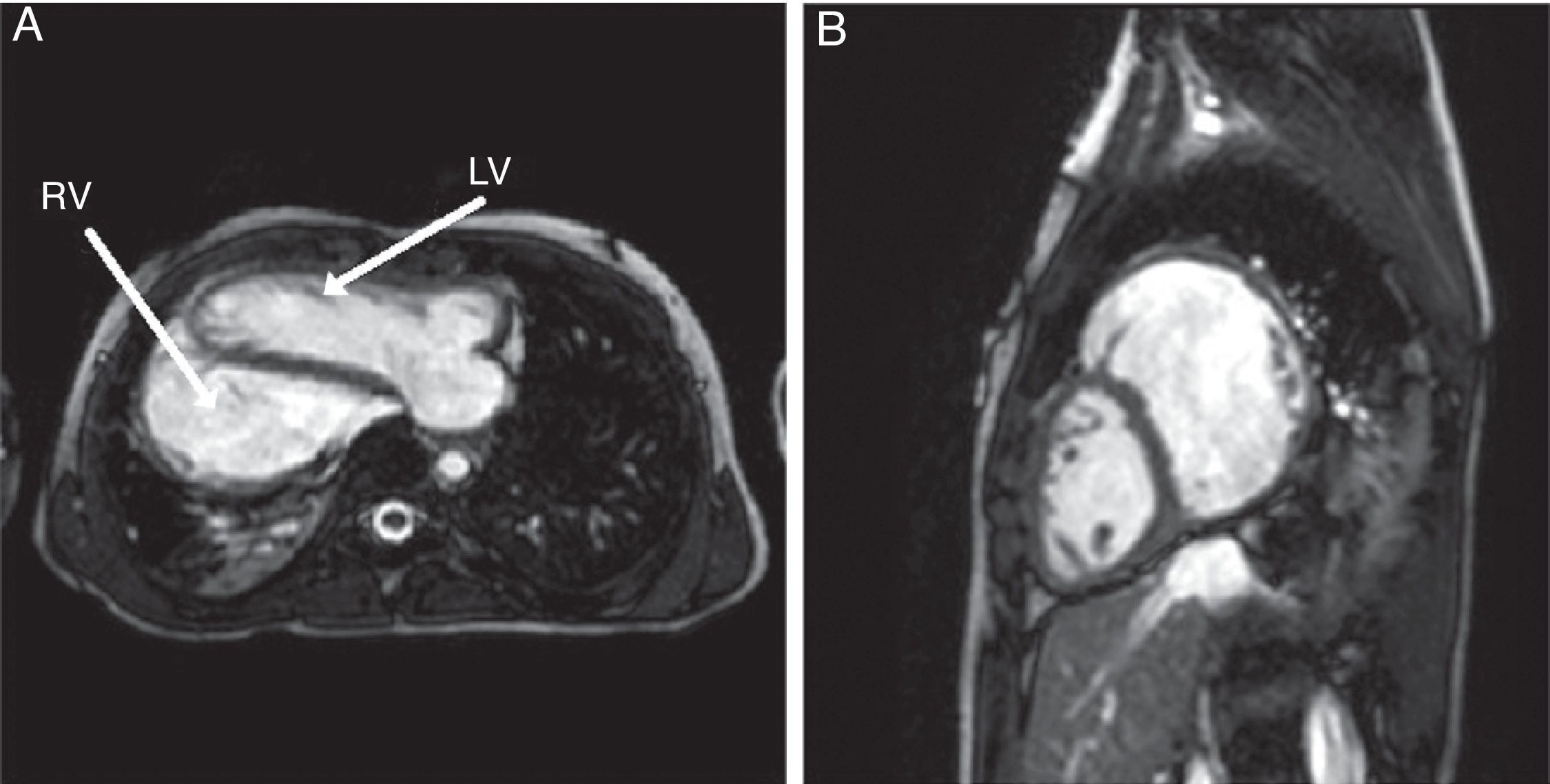
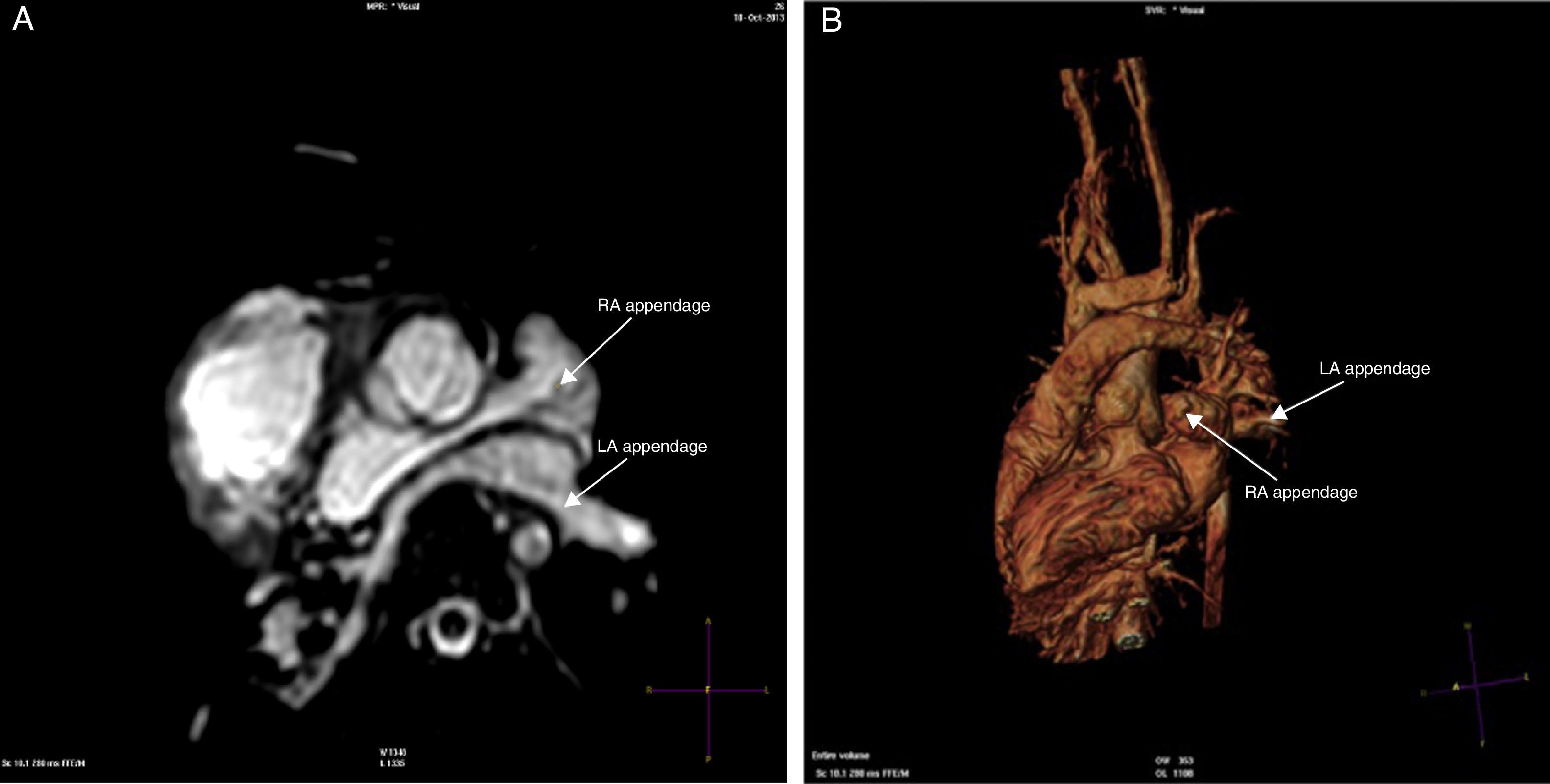
The patient has remained well over the last 12 years. Serial outpatient follow-up revealed pulmonary regurgitation. An MRI scan was performed to quantify the regurgitation, ventricular volumes and function, which showed dextrocardia with normal connections but an unusual ventricular orientation with the right ventricle (RV) to the right and posterior to the left ventricle (LV). The RV was moderately dilated (Figure 1A and B). There was moderate pulmonary regurgitation but no outflow obstruction or branch pulmonary stenosis. The aortic root was mildly dilated. The MRI images clearly delineated juxtaposition of the right atrial appendage (RAA) (left juxtaposition). The RAA is seen coursing posterior to the great arteries towards the left side, over the left atrium (LA), and superior to the left atrial appendage. Both the atrial appendages are seen positioned to the left of the great vessels (Figure 2A and B).
 showing (A) apex to the RV and LV anterior to the RV; (B) LV anterior to the dilated RV.")
 Cardiac MRI (balanced SSFP cine images), axial view, showing juxtaposed RAA on the left side: RAA has triangular shape and LAA has tubular shape. (B) 3D-balanced SSFP volume rendered view showing left juxtaposition.")
Juxtaposition of atrial appendages is a rare congenital cardiac malformation (incidence 1%). Left juxtaposition is six times more frequent than right. The most common anomalies associated with juxtaposition include TGA, tricuspid atresia, cardiac malposition and subaortic infundibulum.
Ethical disclosuresProtection of human and animal subjectsThe authors declare that no experiments were performed on humans or animals for this study.
Confidentiality of dataThe authors declare that they have followed the protocols of their work center on the publication of patient data.
Right to privacy and informed consentThe authors declare that no patient data appear in this article.
Conflicts of interestThe authors have no conflicts of interest to declare.
