
Polysplenia/heterotaxy syndrome is a rare congenital disorder associated with a wide spectrum of anomalies in various organ systems. Although anomalies of the cardiovascular system are common in this syndrome, the authors report a rare case of polysplenia syndrome associated with aortic pseudocoarctation, which to our knowledge has never been reported.
A síndrome de poliesplenia/heterotaxia é uma anomalia congénita rara associado a um amplo espetro de anomalias em vários órgãos. Apesar das anomalias do sistema cardiovascular estarem frequentemente associadas a esta síndrome, os autores relatam um caso raro de uma síndrome poliesplenia associada a uma pseudocoartação aórtica, o que segundo a nosso conhecimento nunca foi descrito na literatura.
Polysplenia syndrome is a rare congenital disorder that is characterized by multiple spleens and duplication of left-sided structures.1–4 A wide spectrum of anomalies of the cardiovascular system, present in up to 75% of cases, is associated with this syndrome.1 However, to our knowledge, pseudocoarctation of the aorta associated with polysplenia syndrome has never been reported. We describe the imaging findings of such a case on multidetector computed tomography (MDCT).
Case reportA 15-year-old boy with symptoms of palpitations and normal physical examination underwent a transthoracic echocardiogram (TTE) that revealed pseudocoarctation and aneurysm of the thoracic aorta, as well as a prominent blood vessel anteriorly crossing the descending aorta, of unknown etiology.
The patient was referred for further investigation by MDCT angiography, which revealed prominent kinking of the aortic arch distal to the origin of the innominate artery and fusiform dilatation of the descending aorta (but with no significant collateral circulation), consistent with the echocardiographic findings of aortic pseudocoarctation (Figure 1a). The prominent blood vessel anteriorly crossing the descending aorta shown on the TTE was identified as a dilated hemiazygos vein (Figure 1b). Bilateral morphological left bronchi were also noted (Figure 1c). A systemic venous drainage anomaly affecting the inferior vena cava (IVC) was suspected and abdominal MDCT imaging was performed. This showed a duplicated IVC with left dominance. The left IVC was formed by the junction of the left internal and external iliac veins and continued cranially through the hemiazygous system without joining the right IVC. The latter was formed by the junction of the left internal and external iliac veins and continued cranially up to the liver, draining into the right suprahepatic vein (Figure 2a). Before continuing as the IVC on both sides, the common iliac veins were interconnected by transverse venous anastomoses and on the left side two renal veins were found draining into the left IVC. An extensively septated left spleen was also present (Figure 2b). Moreover, agenesis of the pancreatic tail and lateral part of the body were also noted, as well as right renal agenesis and left hydronephrosis and hydroureter (Figure 2c and 2d).
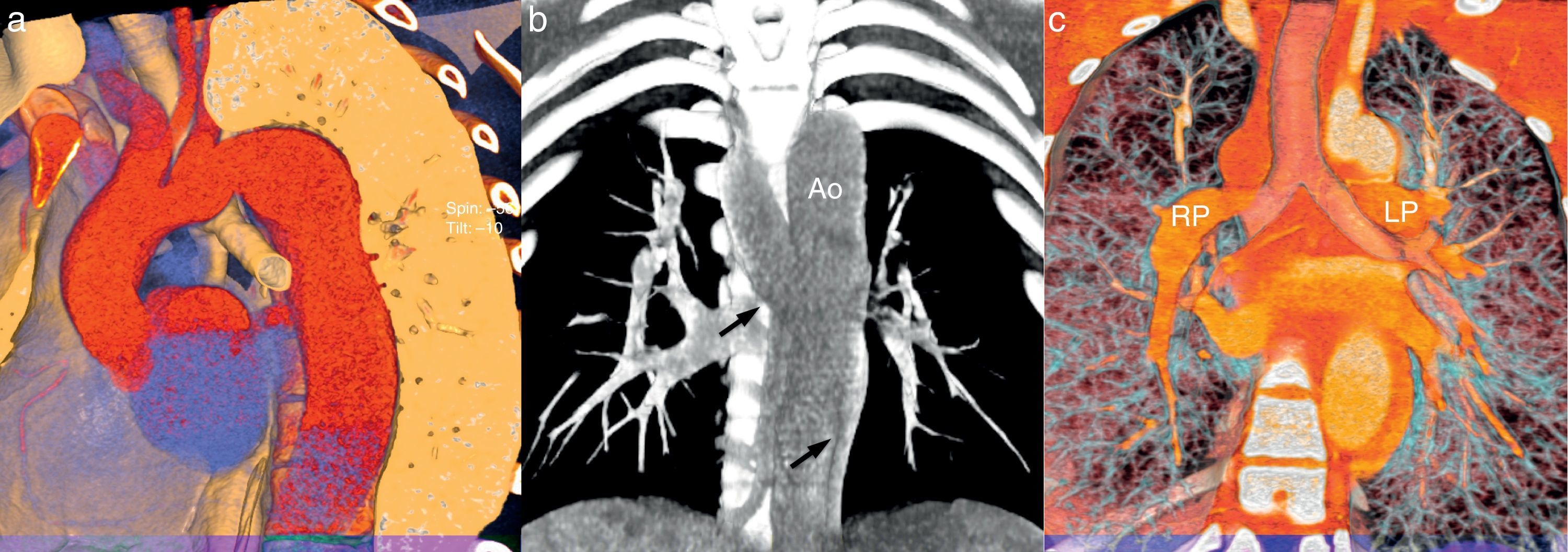
 Three-dimensional volume rendering of the thoracic aorta. The ascending aorta is normal, but there is significant tortuosity of the aortic arch and fusiform dilatation of the descending aorta; (b) coronal plane volume rendering reconstruction showing a dilated hemiazygos vein (black arrows) anteriorly crossing the descending aorta; (c) coronal plane volume rendering reconstruction demonstrating the presence of bilateral left bronchi (hyparterial bronchial position). Ao: aorta; LP: left pulmonary artery; RP: right pulmonary artery.")
(a) Three-dimensional volume rendering of the thoracic aorta. The ascending aorta is normal, but there is significant tortuosity of the aortic arch and fusiform dilatation of the descending aorta; (b) coronal plane volume rendering reconstruction showing a dilated hemiazygos vein (black arrows) anteriorly crossing the descending aorta; (c) coronal plane volume rendering reconstruction demonstrating the presence of bilateral left bronchi (hyparterial bronchial position). Ao: aorta; LP: left pulmonary artery; RP: right pulmonary artery.
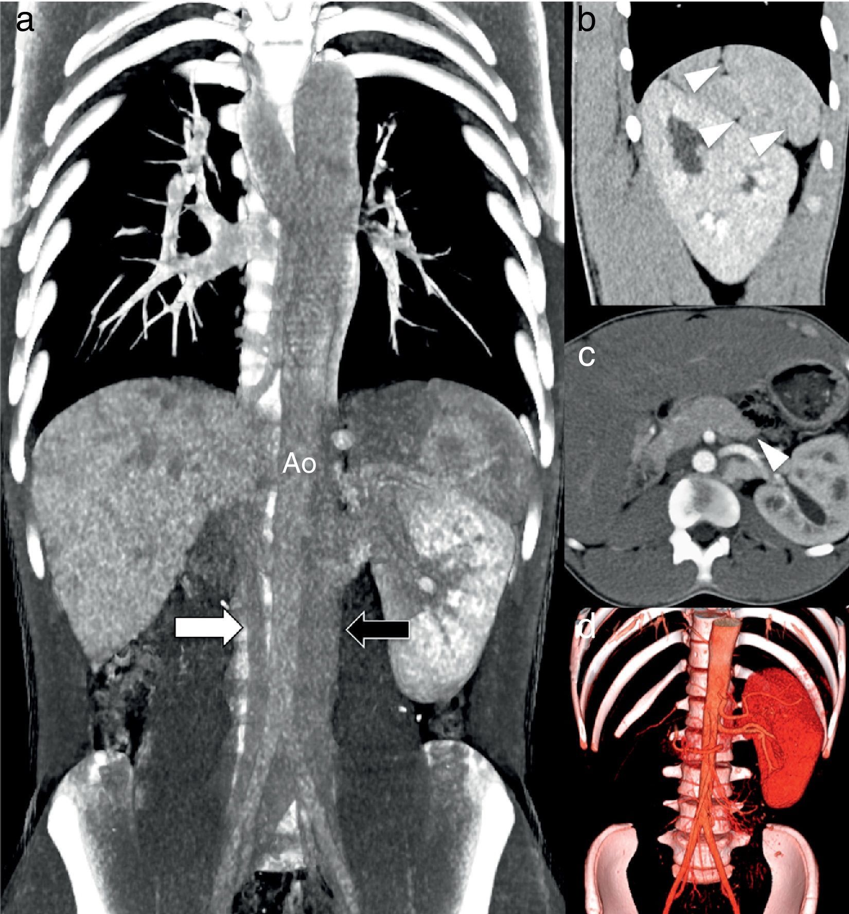
 Coronal plane volume rendering reconstruction showing duplicated IVC with left dominance, with the left IVC (black arrow) continuing cranially through the hemiazygos system without joining the right IVC and the right IVC (white arrow) continuing to the liver; (b) sagittal reformatted contrast-enhanced computed tomography showing an extensively septated spleen consistent with polysplenia (arrows); (c) axial reformatted contrast-enhanced computed tomography showing associated absence of the pancreatic tail and lateral part of the body (arrow); (d) coronal three-dimensional volume rendering showing absence of the right kidney. Ao: aorta.")
(a) Coronal plane volume rendering reconstruction showing duplicated IVC with left dominance, with the left IVC (black arrow) continuing cranially through the hemiazygos system without joining the right IVC and the right IVC (white arrow) continuing to the liver; (b) sagittal reformatted contrast-enhanced computed tomography showing an extensively septated spleen consistent with polysplenia (arrows); (c) axial reformatted contrast-enhanced computed tomography showing associated absence of the pancreatic tail and lateral part of the body (arrow); (d) coronal three-dimensional volume rendering showing absence of the right kidney. Ao: aorta.
These findings were consistent with polysplenia syndrome with heterotaxy.
DiscussionPolysplenia syndrome is characterized by multiple spleens and by a duplication of left-sided structures, such as bilobed lungs, as well as a wide variety of abnormalities in various organ systems.1–4 Typically both lungs have two lobes and hyparterial bronchi, as in the case presented.2
Cardiovascular anomalies are common in this syndrome and are responsible for most deaths in childhood.4 A broad spectrum of cardiovascular defects is associated with polysplenia, including double-outlet right ventricle and left heart obstruction, such as coarctation of the aorta.1,5 In our patient, MDCT showed polysplenia syndrome associated with pseudocoarctation of the aorta, which to our knowledge has never been reported.
Anomalies of systemic venous drainage are also commonly associated, and include interruption of the IVC with azygos continuation and duplication of the IVC.1,6 Our patient showed a duplicated IVC, the left IVC being associated with hemiazygous continuation, while the right IVC entered the right suprahepatic vein.
Anomalies of the pancreas are well known in this syndrome and in most cases a short pancreas with partial agenesis of the dorsal portion is reported, as in our patient.4,5 Although agenesis of the dorsal pancreas generally remains asymptomatic because of the functional reserves of the exocrine and endocrine pancreas, a possible relationship with early- or late-onset diabetes, and with an increased incidence of pancreatitis, has been reported in the literature.4 Since our patient had no clinical endocrine or exocrine pancreatic dysfunction, no further studies were carried out.
Further abdominal anomalies have been reported, include symmetric or midline liver, abnormalities of the biliary tract or gallbladder, such as biliary atresia, malrotation of the bowel and renal agenesis.1–6The interest of this case lies in the novel association between polysplenia/heterotaxy syndrome and aortic pseudocoarctation.
Ethical disclosuresProtection of human and animal subjectsThe authors declare that no experiments were performed on humans or animals for this study.
Confidentiality of dataThe authors declare that no patient data appear in this article.
Right to privacy and informed consentThe authors declare that no patient data appear in this article.
Conflicts of interestThe authors have no conflicts of interest to declare.
