
The authors present the case of a 47-year-old man, with hypertension and dyslipidemia as cardiovascular risk factors, who had come to the emergency department in 2008 for chest pain. Diagnostic exams at that time (ECG and cardiac biomarkers) revealed no signs of acute ischemia, but exercise testing was positive for myocardial ischemia. Invasive coronary angiography was performed, which showed no angiographically significant coronary lesions. He subsequently underwent myocardial perfusion scintigraphy, which revealed no evidence of ischemia.
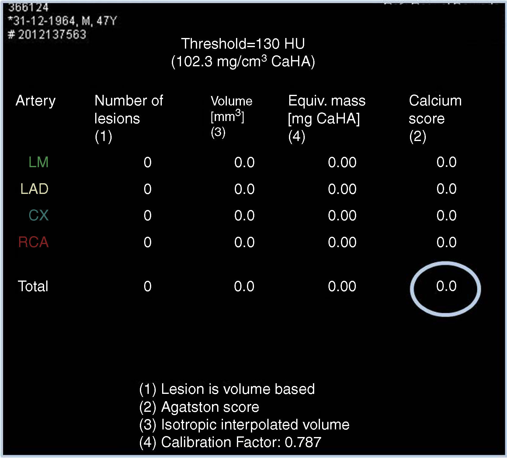
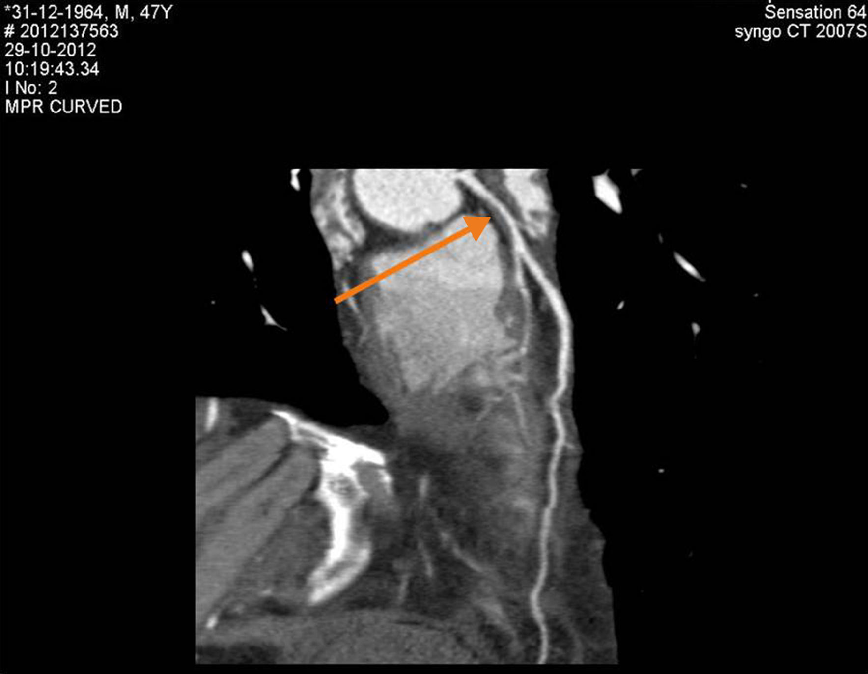
Due to persistence of symptoms, in 2012 exercise testing was repeated, which was positive for ischemia. In order to clarify the clinical picture, he was referred to our hospital for cardiac computed tomography (CT) angiography. This showed a zero calcium score (Figure 1), corresponding to the 25th percentile for age and gender, and revealed 50–70% stenosis with a non-calcified eccentric plaque in the distal left main/proximal anterior descending artery (Figure 2).


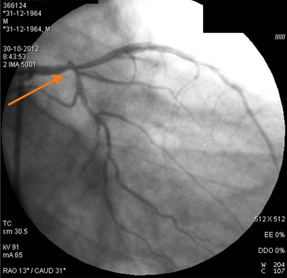
Invasive coronary angiography confirmed significant coronary artery disease (Figure 3) and the patient was referred for coronary artery bypass grafting. The authors did not have access to the initial coronary angiography since this was performed in a different institution, and at referral the patient was in possession of clinical information only, together with the request for cardiac CT angiography.

This case highlights the important contribution that cardiac CT angiography can make to the diagnosis and characterization of coronary artery disease.
Ethical disclosuresProtection of human and animal subjectsThe authors declare that no experiments were performed on humans or animals for this study.
Confidentiality of dataThe authors declare that they have followed the protocols of their work center on the publication of patient data.
Right to privacy and informed consentThe authors declare that no patient data appear in this article.
Conflicts of interestThe authors have no conflicts of interest to declare.
Please cite this article as: Dourado R, Couto R, Pacheco M, Tavares A, Martins D. Contributo da angio tomografia computorizada cardíaca na avaliação da dor torácica. Rev Port Cardiol. 2014;33:653–654.
