

Acute heart failure (AHF) presents significant clinical and economic challenges that require a comprehensive, evidence-based strategy extending from in-hospital management to post-discharge care. Patients with AHF usually require prompt initiation or intensification of HF-directed treatment, which commonly includes intravenous diuretics, and frequently culminates in unplanned hospitalization. Despite therapeutic advances, AHF is still associated with poor outcomes, with in-hospital mortality in Portugal reaching 12.4%. Based on the expertise and clinical experience of a panel of cardiology and internal medicine specialists, this paper provides a practical guide to AHF management over the patient journey, which has been adapted to align with the specificities of the Portuguese national healthcare system: hospital admission, initial management of AHF, in-hospital management of stable phase, pre-discharge and transition of care. This practical guide highlights the importance of optimizing guideline-directed medical therapy by tailoring and rapidly uptitrating treatments to reduce mortality and readmissions. It is equally essential that structured, multidisciplinary transition-of-care programs are established that promote continuity and coordination across care settings.
A insuficiência cardíaca aguda (ICA) apresenta desafios clínicos e económicos significativos que exigem uma estratégia holística e baseada na evidência, que vai desde a gestão hospitalar até aos cuidados pós-alta. Os doentes com ICA requerem geralmente o início imediato ou a intensificação do tratamento dirigido à IC (insuficiência cardíaca), que na maioria dos casos inclui diuréticos intravenosos, e frequentemente conduz à hospitalização não planeada. Apesar dos avanços no tratamento da IC crónica, a ICA está ainda associada a um prognóstico desfavorável, com taxas de mortalidade hospitalar em Portugal que alcançam os 12,4%. Com base na expertise e experiência clínica de um painel de especialistas em Cardiologia e Medicina Interna, neste artigo fornecemos um guia prático para a gestão da ICA ao longo da jornada do doente, adaptado para estar alinhado com especificidades do sistema nacional de saúde português: internamento hospitalar, gestão inicial da ICA, gestão hospitalar da fase estável, pré-alta e transição de cuidados. Este guia prático destaca a importância de otimizar a terapêutica guiada por recomendações clínicas, adaptando e titulando rapidamente os fármacos para reduzir a mortalidade e as readmissões. É igualmente essencial a implementação de programas de transição estruturados e multidisciplinares que garantam a coordenação e continuidade do tratamento.
Acute heart failure (AHF) is characterized by new onset or worsening of symptoms and/or signs of heart failure (HF) requiring urgent medical assessment and treatment.1–3 Patients presenting with AHF usually require immediate initiation or intensification of HF-directed treatment, which commonly includes intravenous (IV) diuretics, and frequently leads to unplanned hospitalization.2,4,5
Despite advances in treatment, AHF continues to carry a poor prognosis. In-hospital mortality ranges from 4 to 10%, rising to as high as 50% in cases of cardiogenic shock. After discharge, overall mortality remains high, with rates of 25–30% within the first year, particularly in the first three months, a period commonly referred to as the “vulnerable phase”.6,7
In Portugal, data from national registries of the Portuguese Directorate-General of Health indicate that in 2016, 18752 patients were hospitalized for HF, and in-hospital mortality stands at 12.4%.8 Additionally, a 2017 study involving 429 patients, reported in-hospital mortality at 7.9%, while at one-year follow-up, mortality and rehospitalization were 34.3% and 30.5%, respectively.9 Portuguese data are comparable with those from other European countries. In a study covering 1995 to 2014, Fonseca et al.10 encountered AHF in-hospital mortality of 5.5–14.0% in Portuguese studies, vs. 3.8–12.0% in studies from Europe.
Drawing on the expertise and clinical experience of a multidisciplinary panel of cardiology and internal medicine specialists, this paper presents a practical, context-specific guide to the management of AHF throughout the patient journey – from hospital admission to transition of care and outpatient follow-up – adapted to the Portuguese healthcare setting. It is meant to build upon and complement the guidelines from the European Society of Cardiology – Heart Failure Guidelines,1,11 from a more practical perspective, considering the specificities of HF care in Portugal.
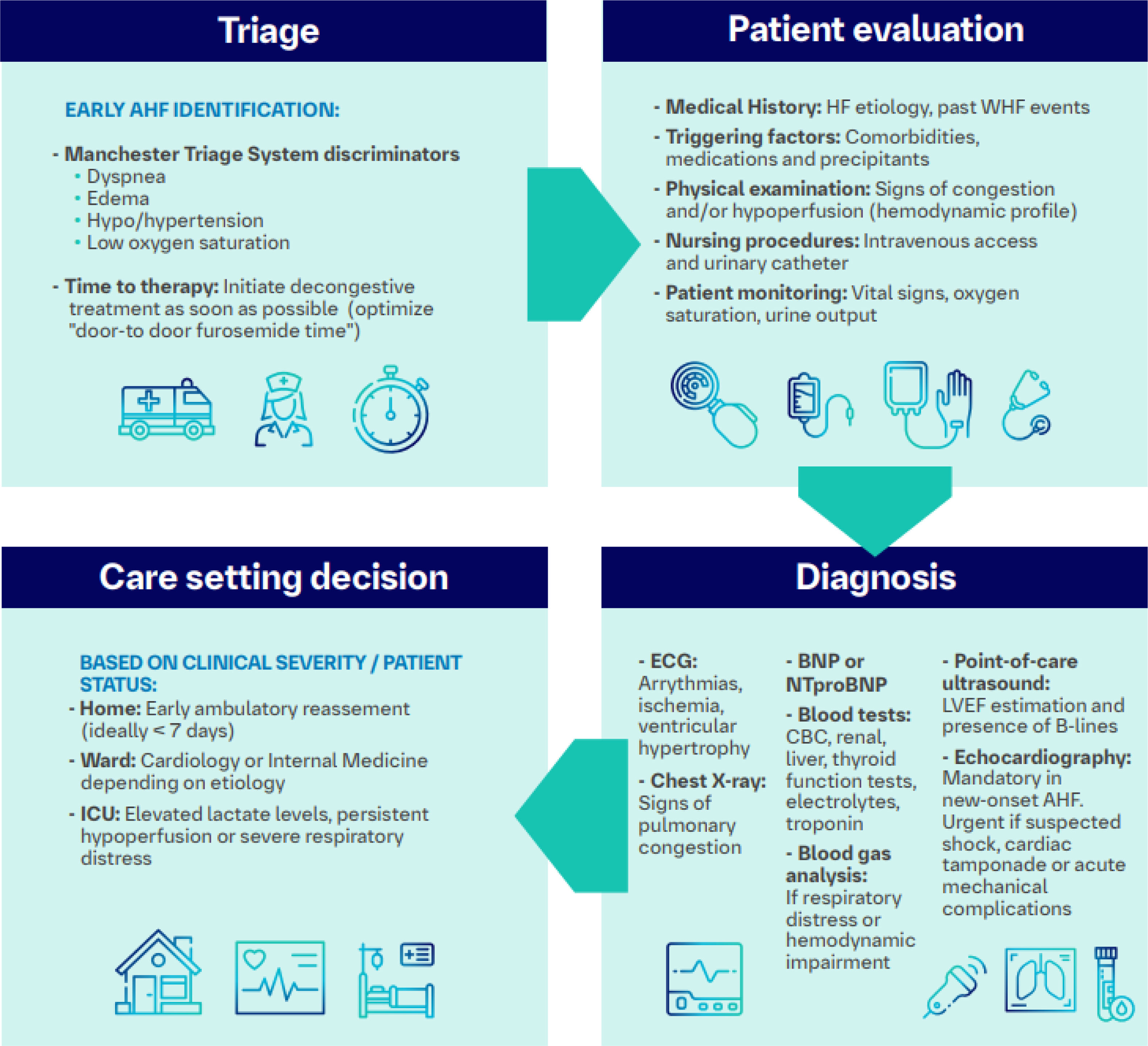
AdmissionAcute HF can present as a clinical deterioration in patients with a prior diagnosis of HF (i.e., acutely decompensated HF) or new onset AHF in patients without known HF (i.e., de novo HF). The clinical presentation of AHF can vary widely depending on the patient's clinical profile, comorbidities, and the underlying etiology.2,3 The evaluation of patients with AHF frequently starts in the emergency room (ER), where evidence-based strategies are limited. Therefore, the clinical approach to AHF varies widely among healthcare professionals and across centers. This section aims to establish a standardized and comprehensive approach to the initial evaluation of patients with AHF in the Portuguese ER context (Figure 1).

AHF admission roadmap. AHF: acute heart failure; BNP: B-type natriuretic peptide; CBC: complete blood count; ECG: electrocardiogram; HF: heart failure; ICU: intensive care unit; LVEF: left ventricular ejection fraction; NTproBNP: N-terminal pro B-type natriuretic peptide; WHF: worsening heart failure.
The primary objective at the time of admission is to establish a diagnosis of AHF rapidly, determine the patient's clinical profile, identify any acute causes or precipitating factors, and initiate appropriate therapy without delay.
TriageVarious clinical studies underpin the importance of a timely diagnosis and rapid IV diuretic initiation (“door-to-furosemide time”).12,13 For instance, shorter intervals between ER arrival and loop diuretic administration are linked to better symptom relief and lower in-hospital mortality.14
Although a structured approach using the Manchester Triage System (MTS) discriminators such as dyspnea, edema, hyper/hypotension and low oxygen saturation may theoretically facilitate the prompt identification and treatment of AHF patients, it lacks a specific flowchart for suspected AHF.15
As the MTS is based on signs and symptoms presented by the patient rather than medical diagnosis, several studies have reported sensitivity for AHF ranging from 44% to 80%.15,16 Consequently, a considerable proportion of patients with AHF may be undertriaged to a lower priority level. To address this limitation, we propose the development and implementation of a dedicated heart failure fast track system that could streamline the triage process and improve outcomes for AHF patients. This service could rely on rapid, accessible discriminators, such as dyspnea, known HF and high point of care natriuretic peptide levels, with quick referral to dedicated inpatient or outpatient HF units/clinics.
General evaluation of the patientThe evaluation of patients should include past medical history, previous HF information such as etiology, left ventricular ejection fraction (LVEF], past worsening events/hospitalizations, comorbidities, and medications. It should try to clarify triggering factors and possible corrections (e.g., CHAMPIT).1 The physical examination should place special emphasis on establishing the clinical-hemodynamic profile based on congestive and hypoperfusion signs.
DiagnosisUpon admission, for patients who have not previously been diagnosed with HF, a de novo HF diagnosis should be established according to the universal definition of HF by integrating clinical presentation, medical history, and relevant diagnostic tests.2 For patients who have already been diagnosed with HF, if acute decompensation is present, a diagnosis of worsening HF should be considered.1
Several conditions, however, can mimic or exacerbate AHF, potentially complicating the diagnosis. Worsening kidney disease, noncardiogenic pulmonary edema, chronic venous insufficiency, obesity, liver disease, thyroid abnormalities, anemia, or chronic obstructive pulmonary disease are some of the conditions that present with overlapping clinical features.17,18
Recognizing and distinguishing these conditions from true AHF is crucial to ensuring accurate diagnosis and timely initiation of targeted therapy. Supplementary Table S1 provides a proposed checklist for the initial evaluation of AHF.
First-line examsFirst-line exams are usually carried out in the emergency department or within the first days of hospitalization and include the following.
ElectrocardiogramAn electrocardiogram (ECG) is essential to assess arrhythmias, ischemia or myocardial infarction, ventricular hypertrophy, and conduction abnormalities that may require targeted treatment. Although a normal electrocardiogram is unlikely in AHF, it is present in 10–15% of AHF patients. Notably, a normal ECG significantly decreases the likelihood of AHF but should not be used to rule out the diagnosis.1,19
Natriuretic peptidesBoth BNP (B-type natriuretic peptide) and NT-proBNP (N-terminal pro B-type natriuretic peptide) levels are important tests to assist in diagnosis and prognosis. In view of the high negative predictive value of natriuretic peptides, normal values upon hospital admission (BNP <100 pg/mL and NT-proBNP <300 pg/mL), especially in patients not on diuretics, make the diagnosis of AHF unlikely.1
Laboratory testsOther blood tests may help assess decompensation factors and should include hemogram, renal, liver, thyroid function tests, electrolytes, and troponin. D-dimers can be helpful when pulmonary thromboembolism is suspected, and C-reactive protein and procalcitonin when infection is suspected.1 Blood gas analysis should be performed in case of respiratory distress or hemodynamic impairment.1 Specific laboratory tests should be tailored to each patient's clinical presentation as they may lead to a confounded diagnosis. For instance, troponin measurement may not be necessary in the absence of clinical features suggestive of acute coronary syndrome, such as chest pain, significant ventricular dysrhythmias, or ECG changes.
Chest X-rayA chest X-ray should be performed early upon admission to help identify pulmonary congestion, pleural and/or pericardial effusion. It also enables differential diagnosis in the case of lung diseases. Computed tomography may be indicated, especially in cases of suspected pulmonary thromboembolism.1
Point of care ultrasoundA point of care ultrasound (POCUS) is a valuable diagnostic tool, increasingly utilized in acute care settings. It enables the identification of pulmonary and systemic congestion and estimation of left ventricle systolic function.20,21
EchocardiogramConfirmation of the diagnosis by echocardiogram is mandatory in new onset AHF and should be performed as soon as possible.2 Nevertheless, it is seldom available for most of the patients during the baseline evaluation in the emergency department and is only performed in the ward during a hospital stay. Some situations, however, require urgent echocardiographic evaluation, such as shock, suspicion of cardiac tamponade, or acute mechanical complications.
Care setting decisionFollowing the initial diagnostic workup, the decision on patient allocation should be guided by the patient's clinical severity, hemodynamic stability, and underlying etiology. Critically unstable patients, those with cardiogenic shock, severe respiratory compromise, or showing signs of end-organ hypoperfusion, normally require admission to an intensive care unit for close monitoring and advanced therapies.1,22 Patients with severe congestion or unknown HF etiology often benefit from admission to a specialized medical ward, where further investigations can clarify the HF etiology.2 Most of those who are clinically stable upon evaluation in the ER are admitted to a general medical ward. Some may be considered for discharge, provided they have early outpatient follow-up arranged, ideally in a dedicated HF clinic, to ensure prompt reassessment and therapy optimization.17 Establishing an etiological diagnosis is particularly important in de novo AHF, as targeted management strategies may differ significantly based on the underlying cause.1
Initial management of AHFAcute heart failure can present under different clinical phenotypes, and initial management depends on the severity of congestion and the presence of hypoperfusion.23 Defining the hemodynamic profile is crucial to guiding initial treatment effectively. Oxygen therapy and non-invasive ventilation are essential measures in patients with low peripheral oxygen saturation levels and significant breathing effort.3
Congestion and hypoperfusion: clinical, laboratory, and imaging evaluationClinical assessment of congestion and hypoperfusionSigns and symptoms of congestion should be systematically assessed.13 Dyspnea at rest or on exertion and orthopnea correlate with elevated left-sided filling pressures, particularly pulmonary capillary wedge pressure, which indicates lung congestion.24 Pulmonary rales, peripheral edema, and jugular vein distention are common signs of congestion.24 Classic signs of hypoperfusion, such as cold sweated extremities, prolonged capillary refill time, oliguria, and confusion, contribute to defining the severity of the clinical presentation but not to the degree of congestion.1 Hypotension and hypoperfusion often coexist, but they are not synonymous. Treatment decisions, therefore, should not rely solely on systolic blood pressure (SBP) levels but also consider markers of end-organ dysfunction and overall perfusion status.13,25
Laboratory and imaging toolsCongestion is usually associated with elevated natriuretic peptides.13 A decrease in NT-proBNP concentration greater than 30% during hospitalization is associated with a better prognosis and more effective decongestion in AHF patients.26,27
A point of care ultrasound has become a valuable tool in the evaluation of AHF.28 The main diagnostic findings include hyperechoic vertical artifacts (B-lines), the number of which suggests the degree of interstitial edema and pleural effusion.29,30 Inferior vena cava (IVC) diameter and respiratory variation are related to the degree of congestion.31 The venous excess ultrasound (VExUS) score, which includes a Doppler evaluation of the IVC, hepatic and portal veins, and intrarenal venous flow, has been gaining more attention for assessing venous congestion.28,32 By detecting early signs of venous congestion, VExUS scoring can help clinicians refine hemodynamic management and potentially improve outcomes, particularly in acutely decompensated patients.33,34
Management of the congestive phase in heart failureUp to 90% of AHF patients show congestion signs at admission, and over one-third remain congestive at discharge.13 Residual congestion at hospital discharge has a clear negative prognostic impact on AHF patients; therefore, during hospitalization, from admission to discharge, all efforts are required to achieve an effective decongestion.13
Diuretic therapy: route, starting dose and monitoringIn congested patients, IV diuretics are required to achieve an effective decongestion as gastroenteric congestion compromises the absorption of oral diuretics. Intravenous furosemide offers reliable bioavailability and a predictable titration dose. Figure 2 presents a management proposal for diuretic therapy in congested patients.

Diuretic therapy. *Urine output <100–150 mL/h within the first 6 hours or urinary Na concentration <50 mEq/L within 2 hours post-administration or worsening of symptoms and signs of congestion; **Same as above+progression from clinical profile B to C (hypoperfusion arising). ASAP: as soon as possible; ER: emergency room; GDMT: guideline-directed medical therapy; HF: heart failure; IV: intravenous; MRA: mineralocorticoid receptor antagonists; RRT: renal replacement therapy; SGLT2i: sodium-glucose co-transporter-2 inhibitors.
The DOSE study revealed that a high-dose of IV furosemide (2.5 times daily oral dose) provided faster and more effective decongestion than low-dose (equal to daily oral dose), but with a transient and reversible increase in serum creatinine.35 There was no difference between bolus (twice a day) and continuous administration.35,36 Nevertheless, continuous infusion may be preferred for higher doses of loop diuretic but requires a loading dose to achieve therapeutic plasma concentrations rapidly.37 As loop diuretics induce diuresis and natriuresis, a diuretic response should be assessed using urine volume and sodium (UNa) concentrations. A satisfactory diuretic response is indicated by achieving a urine output >100–150 mL/h within the first 6 hours or detecting a UNa concentration >50–70 mEq/L within 2 hours post-administration.1 If those thresholds are not met, the diuretic dose should be doubled to attain adequate decongestion.24
Overcoming diuretic resistancePatients with chronic HF and on long-term diuretic therapy may present an impaired sensitivity to diuretics, defined as diuretic resistance.38 Diuretic resistance should be considered in the absence of an effective diuretic response to a high-dose IV loop diuretic therapy (Figure 2).
Strategies to overcome diuretic resistance include:
- 1.
Increasing furosemide dose, by doubling it, up to 400–600 mg/day;
- 2.
Combining furosemide with other agents targeting different nephron sites, known as “sequential nephron blockade”: In the ADVOR trial, acetazolamide, a carbonic anhydrase inhibitor that reduces proximal tubular sodium reabsorption, was found to improve decongestion rates within 72 hours in combination with furosemide.39 Hydrochlorothiazide or metolazone, targeting the distal tubule, enhance natriuresis but may increase risks of kidney dysfunction and electrolyte disturbances (hypokalemia and hyponatremia), as seen in the CLOROTIC and DEA-HF studies.40,41 Metolazone has traditionally been used as the second diuretic in Portugal since access to IV acetazolamide and isolated hydrochlorothiazide is limited.
- 3.
Sodium-glucose cotransporter-2 inhibitors are also current options to overcome diuretic resistance. Their use in hospitalized AHF patients enables better decongestion and guideline-directed medical therapy (GDMT) optimization at the same time.42 These agents, such as empagliflozin (EMPULSE study) and dapagliflozin (DICTATE-AHF study), enhance diuresis and natriuresis with fewer adverse renal and electrolyte effects compared to thiazide diuretics.42–44
Renal function evaluation during AHF hospitalization should not rely exclusively on changes in glomerular function, but also on the interpretation of the tubular response to the diuretic therapy (diuretic response/efficacy) and the capacity to eliminate residual congestion. When creatinine elevation accompanies an adequate decongestion and the introduction or titration of neurohumoral blockade, it should not be associated with a worse prognosis and should not lead to the immediate suspension of the diuretic therapy if congestion persists. This situation is referred to as “pseudo-worsening kidney function”.45,46 On the other hand, extreme variations in creatinine should alert physicians to the development of “true-worsening kidney function”, especially when accompanied by other metabolic disturbances like hyperkalemia or metabolic acidosis.46 In hemodynamically stable patients, strategies such as ‘sequential nephron blockade’ or even ultrafiltration, should be considered, whereas in hemodynamically unstable patients, admission to intensive care units for inotropic, vasopressor, or mechanical circulatory support may be warranted.46
Transition from intravenous diuretics to oral diureticsThe decision to switch from IV to oral diuretics should be guided by evidence of effective decongestion. During hospitalization, clinical improvement, NT-proBNP reduction >30%, and regression of pulmonary B-lines on POCUS correlate with adequate decongestion and should be integrated to avoid subclinical congestion.27,46 Gradual tapering of IV diuretics and ensuring clinical stability upon transition to oral diuretics during 24–48 hours before discharge are recommended.46
Hypoperfusion and use of inotropes and/or vasopressorsAcute heart failure patients presenting with hypoperfusion and congestion, a “wet and cold” hemodynamic profile, may require inotropes or vasopressors.17,22 Hypoperfusion can occur in normotensive or even hypertensive patients and is suggested by signs such as altered consciousness or obtundation; oliguria or anuria; cold extremities or prolonged capillary refill time; elevated lactate levels or low cardiac output confirmed via echocardiography.1,47
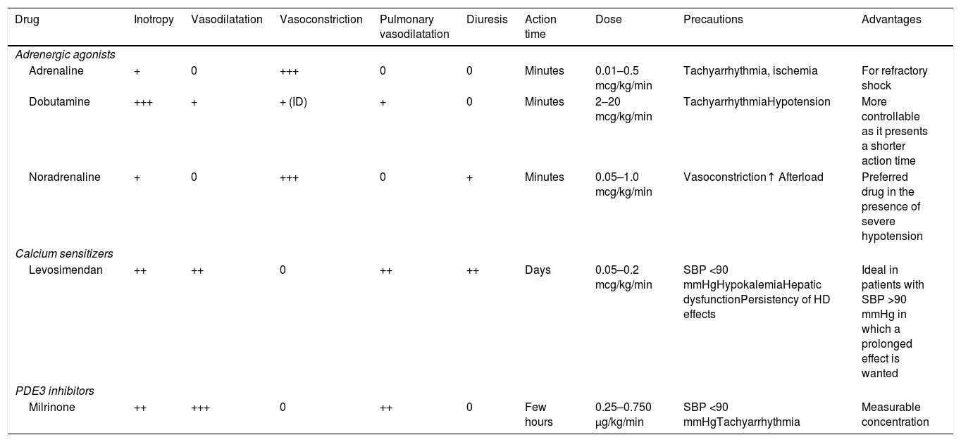
The most used inotropes are dobutamine and levosimendan (Table 1). Dobutamine is a beta-adrenergic agonist with mild inotropic and vasodilatory properties. Common side effects of its use include tachycardia, arrhythmias, and ischemia. Levosimendan enhances troponin's sensitivity to calcium, providing inotropic and vasodilatory effects. Unlike dobutamine, it does not cause tachyphylaxis and has prolonged action due to its metabolites (9–14 days). However, potential adverse events include hypotension and arrhythmias.48,49 Milrinone, a PDE3 inhibitor, also exhibits positive inotropic and vasodilatory properties, but it can cause similar adverse effects. Dopamine use is discouraged due to higher mortality risks associated with arrhythmic causes.50
Inotropic/vasopressor therapeutics.
| Drug | Inotropy | Vasodilatation | Vasoconstriction | Pulmonary vasodilatation | Diuresis | Action time | Dose | Precautions | Advantages |
|---|---|---|---|---|---|---|---|---|---|
| Adrenergic agonists | |||||||||
| Adrenaline | + | 0 | +++ | 0 | 0 | Minutes | 0.01–0.5 mcg/kg/min | Tachyarrhythmia, ischemia | For refractory shock |
| Dobutamine | +++ | + | + (ID) | + | 0 | Minutes | 2–20 mcg/kg/min | TachyarrhythmiaHypotension | More controllable as it presents a shorter action time |
| Noradrenaline | + | 0 | +++ | 0 | + | Minutes | 0.05–1.0 mcg/kg/min | Vasoconstriction↑ Afterload | Preferred drug in the presence of severe hypotension |
| Calcium sensitizers | |||||||||
| Levosimendan | ++ | ++ | 0 | ++ | ++ | Days | 0.05–0.2 mcg/kg/min | SBP <90 mmHgHypokalemiaHepatic dysfunctionPersistency of HD effects | Ideal in patients with SBP >90 mmHg in which a prolonged effect is wanted |
| PDE3 inhibitors | |||||||||
| Milrinone | ++ | +++ | 0 | ++ | 0 | Few hours | 0.25–0.750 μg/kg/min | SBP <90 mmHgTachyarrhythmia | Measurable concentration |
HD: hemodynamic; ID: increased dose; SBP: systolic blood pressure.
In patients with severe hypotension, vasopressors may be necessary to maintain organ perfusion.17 Noradrenaline is the preferred option due to its relative safety concerning arrhythmias.1,17 Inotropes and vasopressors can be used together51. In patients with severe and persistent symptoms, it is important to identify those with criteria for advanced HF early for referral to appropriate centers (i.e., centers capable of providing advanced HF therapies).1
In-hospital management of stable phaseDuring the stable phase, the goals are to confirm or reassess HF etiology, control comorbidities, and start or optimize evidence-based disease-modifying therapy.1
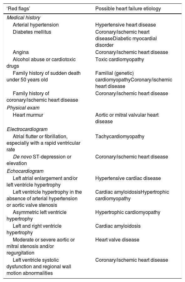
Etiology of heart failureAs stated before, complementary diagnostic tests are essential throughout the entire course of AHF management. New-onset AHF or atypical presentations should be discussed in multidisciplinary teams because these cases may require invasive tests, advanced imaging methods, or genetic testing. Table 2 presents frequently encountered “red flags” and the corresponding possible underlying HF etiology. Confirmation of the diagnosis by echocardiogram is mandatory and should be conducted as soon as possible following suspected AHF. It provides information on functional and structural cardiac abnormalities, enabling evaluation of the LVEF and right ventricular function, regional wall motion abnormalities, valve function, signs of diastolic dysfunction, presence of pulmonary hypertension and pericardial disease. Based on the initial findings and clinical suspicion, additional tests may be necessary to determine or confirm the HF etiology.
‘Red flags’ and possible underlying heart failure etiology.
| ‘Red flags’ | Possible heart failure etiology |
|---|---|
| Medical history | |
| Arterial hypertension | Hypertensive heart disease |
| Diabetes mellitus | Coronary/ischemic heart diseaseDiabetic myocardial disorder |
| Angina | Coronary/ischemic heart disease |
| Alcohol abuse or cardiotoxic drugs | Toxic cardiomyopathy |
| Family history of sudden death under 50 years old | Familial (genetic) cardiomyopathyCoronary/ischemic heart disease |
| Family history of coronary/ischemic heart disease | Coronary/ischemic heart disease |
| Physical exam | |
| Heart murmur | Aortic or mitral valvular heart disease |
| Electrocardiogram | |
| Atrial flutter or fibrillation, especially with a rapid ventricular rate | Tachycardiomyopathy |
| De novo ST-depression or elevation | Coronary/ischemic heart disease |
| Echocardiogram | |
| Left atrial enlargement and/or left ventricle hypertrophy | Hypertensive cardiac disease |
| Left ventricle hypertrophy in the absence of arterial hypertension or aortic valve stenosis | Cardiac amyloidosisHypertrophic cardiomyopathy |
| Asymmetric left ventricle hypertrophy | Hypertrophic cardiomyopathy |
| Left and right ventricle hypertrophy | Cardiac amyloidosis |
| Moderate or severe aortic or mitral stenosis and/or regurgitation | Heart valve disease |
| Left ventricle systolic dysfunction and regional wall motion abnormalities | Coronary/ischemic heart disease |
A transesophageal echocardiogram is recommended for patients with an inconclusive or suspicious transthoracic echocardiogram, especially of valvular diseases, such as endocarditis or mitral and aortic valve disease, and evaluation of intracavitary masses or thrombus, or patients in whom the conditions for undergoing this exam are unfavorable, for example, obesity and patients on mechanical ventilation). Imaging stress tests, such as echocardiography, nuclear stress tests, and cardiac magnetic resonance (CMR), may be indicated for evaluating ischemia or myocardial viability, with the choice depending on accessibility and the experience available at each center. Coronary computed angiogram is indicated to exclude coronary artery disease in patients with new-onset heart failure with reduced ejection fraction (HFrEF) of unclear etiology and low-to-intermediate pre-test probability, and can serve as an anatomical alternative when noninvasive ischemia tests are inconclusive.1 CMR imaging provides a detailed evaluation of cardiac structure, function and tissue characteristics. It plays a crucial role in evaluating cardiomyopathies, helping to enhance diagnosis and guide effective management strategies. Bone scintigraphy, serum free light chain quantification and serum and urine immunofixation are indicated if cardiac amyloidosis is suspected.52
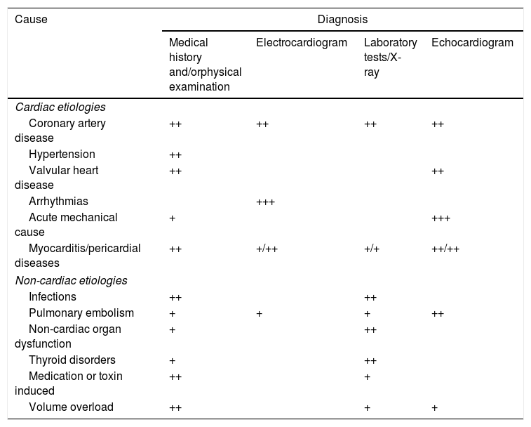
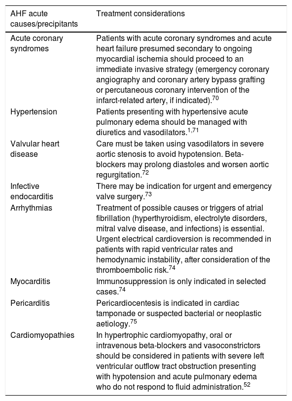
In cases of family history or other indicators of genetic cardiomyopathies, a genetic study should be considered.52 Endomyocardial biopsy can play a crucial role in the evaluation of certain patients with cardiomyopathies or myocarditis, particularly when non-invasive diagnostic methods are inconclusive or when direct histological assessment of myocardial tissue is necessary.53Table 3 outlines the relative importance of diagnostic exams for identifying AHF causes, and Table 4 illustrates the importance of identifying the etiology to guide treatment decisions.
Relative importance of some diagnostic exams for identifying acute heart failure causes.
| Cause | Diagnosis | |||
|---|---|---|---|---|
| Medical history and/orphysical examination | Electrocardiogram | Laboratory tests/X-ray | Echocardiogram | |
| Cardiac etiologies | ||||
| Coronary artery disease | ++ | ++ | ++ | ++ |
| Hypertension | ++ | |||
| Valvular heart disease | ++ | ++ | ||
| Arrhythmias | +++ | |||
| Acute mechanical cause | + | +++ | ||
| Myocarditis/pericardial diseases | ++ | +/++ | +/+ | ++/++ |
| Non-cardiac etiologies | ||||
| Infections | ++ | ++ | ||
| Pulmonary embolism | + | + | + | ++ |
| Non-cardiac organ dysfunction | + | ++ | ||
| Thyroid disorders | + | ++ | ||
| Medication or toxin induced | ++ | + | ||
| Volume overload | ++ | + | + | |
Treatment considerations according to acute causes/precipitants of acute heart failure.
| AHF acute causes/precipitants | Treatment considerations |
|---|---|
| Acute coronary syndromes | Patients with acute coronary syndromes and acute heart failure presumed secondary to ongoing myocardial ischemia should proceed to an immediate invasive strategy (emergency coronary angiography and coronary artery bypass grafting or percutaneous coronary intervention of the infarct-related artery, if indicated).70 |
| Hypertension | Patients presenting with hypertensive acute pulmonary edema should be managed with diuretics and vasodilators.1,71 |
| Valvular heart disease | Care must be taken using vasodilators in severe aortic stenosis to avoid hypotension. Beta-blockers may prolong diastoles and worsen aortic regurgitation.72 |
| Infective endocarditis | There may be indication for urgent and emergency valve surgery.73 |
| Arrhythmias | Treatment of possible causes or triggers of atrial fibrillation (hyperthyroidism, electrolyte disorders, mitral valve disease, and infections) is essential. Urgent electrical cardioversion is recommended in patients with rapid ventricular rates and hemodynamic instability, after consideration of the thromboembolic risk.74 |
| Myocarditis | Immunosuppression is only indicated in selected cases.74 |
| Pericarditis | Pericardiocentesis is indicated in cardiac tamponade or suspected bacterial or neoplastic aetiology.75 |
| Cardiomyopathies | In hypertrophic cardiomyopathy, oral or intravenous beta-blockers and vasoconstrictors should be considered in patients with severe left ventricular outflow tract obstruction presenting with hypotension and acute pulmonary edema who do not respond to fluid administration.52 |
In-hospital care provides a unique opportunity to change the natural history of the disease. This critical period is ideal for optimizing treatment by initiating, adjusting, or titrating prognosis-modifying therapies, thereby reducing the risk of hospital readmission. In HFrEF, GDMT should be gradually uptitrated as tolerated, aiming to implement all four pillars of GDMT. Based on the result from STRONG-HF,54,55 in hemodynamically stable hospitalized AHF patients who respond well to diuretics, early initiation, rapid dose escalation of oral HF therapies, and close follow-up within the first six weeks after discharge is recommended to reduce HF readmissions or all-cause mortality.1 Stability is generally defined as having an SBP of at least 100 mmHg, no use of IV inotropic therapy in the prior 24 hours, and no escalation in IV diuretics or vasodilators within the past six hours.56 Care should be taken to avoid intravascular volume depletion, as this can lead to hypotension and intolerance to renin-angiotensin system antagonists. Angiotensin receptor–neprilysin inhibitors (ARNi) should replace angiotensin converting enzyme inhibitors or angiotensin receptor blockers (ARB).
The TRANSITION study confirmed the safety of initiating sacubitril/valsartan during hospitalization, even for newly diagnosed patients.57 While the PIONEER study showed that early ARNi initiation reduces NT-proBNP and may further lower readmissions.58
Follow-up should prioritize assessing symptoms and signs of congestion, blood pressure, heart rate, NT-proBNP levels, potassium and estimated glomerular filtration rate. For patients with lower blood pressure, starting with a low dose of an ARB may be appropriate, with a potential transition to sacubitril/valsartan before discharge if a low dose of beta-blocker is tolerated (e.g., bisoprolol 1.25 mg once daily or carvedilol 3.125 mg twice daily).56 In cases of mild renal function decline or asymptomatic blood pressure reductions during HF hospitalization, GDMT discontinuation should not be routinely warranted.
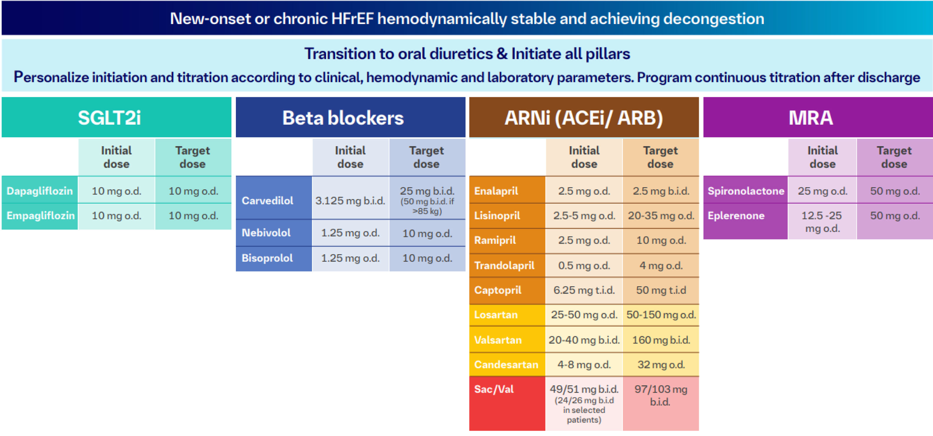
In patients with established HFrEF, if discontinuation of GDMT becomes necessary during hospitalization, it should be restarted promptly as soon as the patient is clinically stable. Multiple studies have highlighted the detrimental effects of interrupting these therapies.59,60 Key aspects of medical therapy in the HFrEF stable phase are presented in Figure 3.

Key aspects of medical therapy in the stable phase. ACEi: angiotensin converting enzyme inhibitors; ARB: angiotensin receptor blockers; ARNi: angiotensin receptor–neprilysin inhibitors; b.i.d.: twice a day; HFrEF: heart failure with reduced ejection fraction; MRA: mineralocorticoid receptor antagonists; o.d.: once a day; Sac/Val: sacubitril/valsartan; SGLT2i: sodium-glucose co-transporter-2 inhibitors; t.i.d.: three times a day.
Although STRONG-HF included patients with HmrEF and HFpEF, evidence supporting use of the four pillars of GDMT in those patients is limited. SGT2 inhibitors have a class I indication irrespective of LVEF class and should, accordingly, be initiated in all HF patients, including in AHF setting.11,42 The other three therapeutic classes may be considered in HFmrEF.1,11 In HF with LVEF>=40%, recent evidence from FINEARTS-HF trial showed that finerenone reduced the composite outcome of CV death and HF events, including in patients who have experienced a recent worsening HF event.61
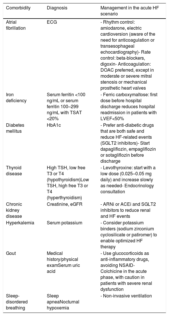
Identification and management of comorbiditiesPatients with HF usually have other cardiovascular and non-cardiovascular diseases that can be the cause of HF, can worsen the prognosis, and frequently make it challenging to optimize HF treatment and use drugs that improve HF prognosis and survival. Table 5 presents the comorbidities most frequently observed in patients with AHF, as well as those for which there is evidence that their management during the AHF episode has a prognostic impact.1
Comorbidities management in the acute heart failure scenario.
| Comorbidity | Diagnosis | Management in the acute HF scenario |
|---|---|---|
| Atrial fibrillation | ECG | - Rhythm control: amiodarone, electric cardioversion (aware of the need for anticoagulation or transesophageal echocardiography)- Rate control: beta-blockers, digoxin- Anticoagulation: DOAC preferred, except in moderate or severe mitral stenosis or mechanical prosthetic heart valves |
| Iron deficiency | Serum ferritin <100 ng/mL or serum ferritin 100–299 ng/mL with TSAT <20% | - Ferric carboxymaltose: first dose before hospital discharge reduces hospital readmission in patients with LVEF<50% |
| Diabetes mellitus | HbA1c | - Prefer anti-diabetic drugs that are both safe and reduce HF-related events (SGLT2 inhibitors)- Start dapagliflozin, empagliflozin or sotagliflozin before discharge |
| Thyroid disease | High TSH, low free T3 or T4 (hypothyroidism)Low TSH, high free T3 or T4 (hyperthyroidism) | - Levothyroxine: start with a low dose (0.025–0.05 mg daily) and increase slowly as needed- Endocrinology consultation |
| Chronic kidney disease | Creatinine, eGFR | - ARNi or ACEi and SGLT2 inhibitors to reduce renal and HF events |
| Hyperkalemia | Serum potassium | - Consider potassium binders (sodium zirconium cyclosilicate or patiromer) to enable optimized HF therapy |
| Gout | Medical history/physical examSerum uric acid | - Use glucocorticoids as anti-inflammatory drugs, avoiding NSAID- Colchicine in the acute phase, with caution in patients with severe renal dysfunction |
| Sleep-disordered breathing | Sleep apneaNocturnal hypoxemia | - Non-invasive ventilation |
ACEi: angiotensin-converting-enzyme inhibitors; ARNi: angiotensin receptor-neprilysin inhibitor; DOAC: direct oral anticoagulants; ECG: electrocardiogram; eGFR: estimated glomerular fraction rate; HbA1c: hemoglobin A1C; HF: heart failure; LVEF: left ventricular ejection fraction; NSAID: nonsteroidal anti-inflammatory drugs; SGLT2: sodium-glucose co-transporter-2; TSAT: transferrin saturation; TSH: thyroid stimulating hormone.
Patients discharged after an AHF episode have high rates of readmission and mortality in the first 2–3 months after hospitalization.62 Although these numbers are not well documented in Portugal, it is estimated that around 25% of patients are readmitted within 30 days and 50% in six months following discharge after an AHF episode.63 Accordingly, interventions targeting this transition period have high potential to impact prognosis and healthcare costs.
Many transition care programs have been developed for AHF patients, with heterogeneous designs, methods and endpoints, leading to inconsistent results. HF management is complex and roadblocks at the patient, doctors, family, hospital, and primary care levels can be encountered.62,64 We believe that the most successful strategies are those tailored to the local context and available resources. The specific characteristics of the Portuguese healthcare system should be taken into account when improving the transition of care among HF patients in Portugal.
Most transition programs (generally coordinated by a HF nurse) include eight common features: patient education, early assessment after admission, medication reconciliation, inclusion of caregivers, telephone follow-up, early follow-up after discharge, home visits, and handoff to post-hospital providers.62 These features can be adapted to the Portuguese context; however, we believe that in-hospital multidisciplinary team discussion, planning and treatment optimization should also be added to the program. As an integrated transition program for implementation in Portugal, we recommend:
- 1.
Admission period:
- •
Early assessment after hospital admission – Transition planning should start at the beginning of hospital admission. An early assessment, usually performed by an HF nurse integrated into an HF team, should include patient and family HF literacy and identify gaps to be corrected before discharge. It also helps with planning home-care services and identifying equipment requirements (e.g., hospital beds, walking aids, home adaptations) needed after discharge, as well as social and/or financial support. This should be done in close collaboration with the social work team, as Portuguese HF patients are often older and socioeconomically disadvantaged.65 Involving key caregivers and family is crucial to optimize HF literacy, treatment adherence, psychosocial support, and monitor early signs of disease decompensation.
- •
In-hospital multidisciplinary assessment: any hospitalized AHF patient's case should be discussed by a HF multidisciplinary team to identify the main issues in each patient, including a full profiling of the HF and comorbidities, identification and management of AHF precipitants and definition of tailored management strategies both during hospitalization and post-discharge.
- •
Treatment optimization: HF treatments should be optimized according to individual patient tolerability, with every effort made to ensure the implementation of prognosis-modifying therapies, as this strategy improves outcomes following an AHF admission.54 Comorbidities should be addressed (section 4.3/Table 5). Indication and referral for advanced HF treatments and palliative care or other therapeutic/diagnostic interventions should be considered. All these domains should be discussed by the HF team and the patients/caregivers while they are still in the hospital and should be clearly written in the discharge letter addressed to the patients, their families and their primary care doctor.
- •
- 2.
Pre-discharge:
- •
Patient education: HF nurses should educate patients when stable before discharge, regarding diet and exercise, HF signs and symptoms monitoring, self-care expectations and strategies, medication education and counselling. This training should address both patients and caregivers. Support materials can be provided.
- •
Medication reconciliation: Redundant and non-evidence-based treatments should be eliminated to simplify treatment regimens while maintaining essential medicines. Treatment adherence is a primary goal and HF nurses and dedicated hospital pharmacists play a crucial role in this step.
- •
Checklist: It is of utmost importance for hospital discharge to take place whenever the patient's condition has been optimized and a safe transition is anticipated. A pre-discharge checklist, such as the one previously developed by the Núcleo de Estudos em Insuficiência Cardíaca (Supplementary Table S2),66 is useful to verify that the most relevant HF issues, comorbidities and organizational matters have been satisfactorily addressed during hospitalization, confirming discharge readiness. Patients in whom any of the identified issues could not be managed should be flagged for a closer follow-up.
- •
Handoff to post-hospital providers: To facilitate multidisciplinary care, a printed discharge summary of the hospitalization is recommended as the official handoff tool. Primary care physicians should have an easy, simple way to communicate with the HF team (e.g., a dedicated phone number written in the handoff letter). Ideally, an alert system could notify primary care doctors whenever their patients are hospitalized/discharged, to facilitate appropriate interactions between hospital and primary care teams, favoring integrated care.
- •
- 3.
Early post-discharge period
- •
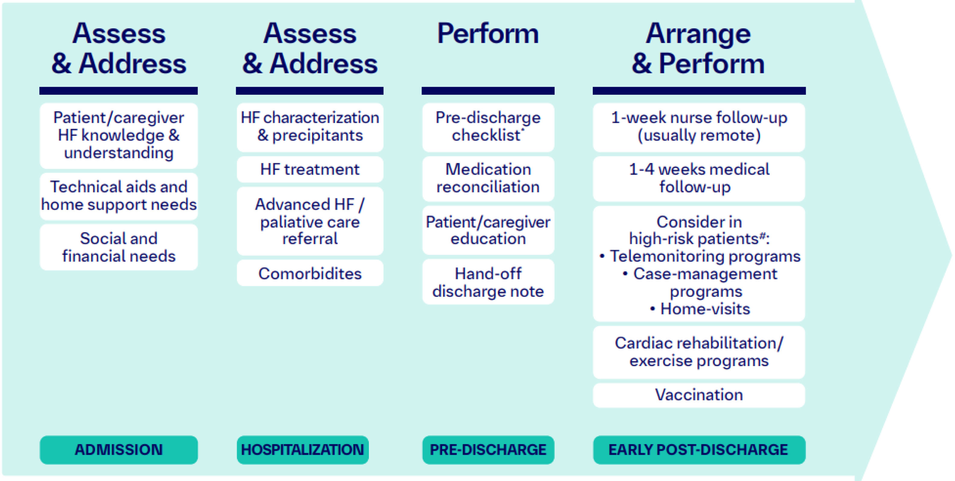
Telephone follow-up: To provide education and support, manage symptoms, and recognize complications early after hospitalization, HF nurses can use post-discharge follow-up telephone calls. We suggest a phone call within one week after discharge, tailoring the frequency of subsequent contact with patient preferences and needs. The identification of patients at high-risk of readmission could support this tailored follow-up planning (Figure 4). A case management strategy could be helpful for chronic complex HF patients, which is already in place at a few Portuguese centers; chronic complex patient units should liaise regarding care with the HF team.
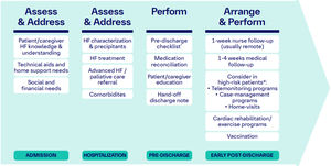 Figure 4.
Transition program for acute heart failure patients. #High-risk patients: patients with ≥1 hospital admission or emergency department visit within 3 months, ≥75 years, with comorbidities (chronic kidney disease [sustained estimated glomerular filtration rate <60 mL/min/1.73 m2 and/or urinary albumin-to-creatinine ratio ≥30 mg/g], chronic obstructive pulmonary disease, cerebrovascular disease, dementia, depression, diabetes or cancer), deficient socio-economic support, high pre-discharge NT-proBNP levels (>1000 pg/mL) and low systolic blood pressure (<100 mmHg) have higher risk for adverse outcomes following an AHF admission. HF: heart failure.
- •
Early follow-up visits after discharge: We suggest at least one medical visit within one to four weeks after hospital discharge, preferably in-person but it can be performed remotely based on patient preferences/limitations. The day hospital is an adequate setting for this re-evaluation. The primary care physician could also carry out this visit. Clinical re-evaluation and medical therapy up-titration are crucial steps in this visit, which should also include other important actions to optimize patient management. In Supplementary Table S3, we propose a checklist for this early post-discharge appointment.
- •
Home visits: This intervention is not ‘mandatory’ but should be implemented whenever possible (by a HF nurse) within the early phase after hospital discharge (one to two months) to reinforce education and help patients in their self-care decisions.
- •
Telemonitoring: In some cases, a remote monitoring approach could be useful. This is already in place at some Portuguese centers and is usually reserved for patients at higher risk of adverse outcomes.
- •
Cardiac rehabilitation: Although not currently available at many Portuguese centers, the integration of a supervised, exercise-based, cardiac rehabilitation program should be considered in patients with more severe disease, frailty, or with comorbidities.1 These rehabilitation programs can be performed in a ‘home-based’ context for patients who live far away from the tertiary center or cannot attend frequent hospital appointments. A detailed implementation of a ‘home-based’ HF rehabilitation strategy in Portugal has been previously reported.67
- •

In short, we believe these steps can be implemented for the transition of care at most tertiary care facilities in Portugal. HF nurses play a vital role in these multidisciplinary programs. We are aware that most centers in Portugal do not have specialized HF nurses; however, institutions should try to identify motivated personnel to set up an HF team with the main goal of managing patients following best practice standards. Ideally, these HF teams should have a minimum of two nurses, two internal medicine specialists, and one cardiologist. These are the minimum human resources expected in centers classified as level 2 under the 2023 National Hospital Referral Network Proposal,68 or as community centers according to the European Society of Cardiology's Quality of Care Centre classification.69 The effectiveness of transition programs should be monitored.
ConclusionAcute heart failure poses significant clinical and economic challenges that require a comprehensive, evidence-based approach extending from in-hospital management to post-discharge care.
This practical guide highlights the importance of optimizing GDMT by tailoring and rapidly up-titrating treatments as needed to reduce mortality and readmissions. The implementation of structured, multidisciplinary transition programs that ensure the continuity of care is equally essential. These programs should incorporate early in-hospital assessments, the use of pre-discharge checklists (e.g., from the NEIC), and robust patient education initiatives led by specialized HF nurses.
Effective AHF management also requires addressing comorbidities, simplifying medication regimens, and engaging both patients and caregivers in self-care strategies. While we acknowledge the constraints faced by many Portuguese centers, this guide advocates for the proactive formation of dedicated HF teams to support best practice standards. Through the adoption of these integrated, patient-focused strategies in local healthcare settings, patient clinical outcomes can be enhanced, the burden of hospital readmissions can be reduced, and the quality of life of patients affected by AHF can be improved.
FundingAna Santos from Prime Focus provided medical writing support, which was supported by Boehringer Ingelheim and Novartis. Boehringer Ingelheim and Novartis had no role in the design, analysis or interpretation of the results in this study. Boehringer Ingelheim was given the opportunity to review the manuscript for medical and scientific accuracy as it relates to Boehringer Ingelheim substances, as well as intellectual property considerations.
Conflicts of interestJoana Pimenta has received speaker/consultant fees from AstraZeneca, Bayer, Bial, Boehringer Ingelheim/Lilly, Daiichi-Sankyo, GSK, Novartis, Pfizer, Servier and Vifor Pharma.
João Pedro Ferreira has no conflicts of interest to declare.
Francisco Vasques-Nóvoa has received consulting or speaker fees from Amgen, AstraZeneca, Bayer, Boehringer Ingelheim, Daiichi Sankyo, Novartis and Ultragenyx.
David Roque has received speaker fees from Bial, AstraZeneca, Novartis, Servier, Boehringer Ingelheim and Orion Pharma.
Inês Araújo has received speaker fees from AstraZeneca, Bayer, Bial, Novartis, Servier, and Advisory fees from AstraZeneca, Bayer, Novartis and Boehringer Ingelheim/Lilly.
César Lourenço has no conflicts of interest to declare.
Susana Dias da Costa has received speaker fees from Boehringer Ingelheim and Novartis Portugal but not related to this paper.
Sara Gonçalves has received speaker fees from AstraZeneca.
Ana Neto has received speaker fees from Novartis, Bial and Daiichi Sankyo.
Filipa Almeida has no conflicts of interest to declare.
Pedro Moraes Sarmento has received consultant or speaking fees from AstraZeneca, Bayer, Boehringer Ingelheim, Lilly and Pfizer.
João Agostinho has received consultant and speaking fees from AstraZeneca, Bayer, Bial, Boehringer Ingelheim & Lilly, Alnylam, Novartis, Daiichi Sankyo and Pfizer.
Irene Marques has received speaker and consultant fees from AstraZeneca, Bayer, Bial, CSL Vifor, Daiichi-Sankyo, Lilly, Novartis, Novo Nordisk, Pfizer, Roche Diagnostics and Servier.
Elisabete Martins has received speaker and consultant fees from AstraZeneca, Bial, Amicus Therapeutics, Sanofi, Amgen and Pfizer.
The following are the supplementary data to this article: