

A 41-year-old woman presented with progressive dyspnea and New York Heart Association functional class II symptoms. Physical examination revealed a grade 3/6 diastolic heart murmur at the lower left sternal border. Transthoracic echocardiography (TTE) showed severe aortic valve regurgitation, a giant right sinus of Valsalva aneurysm (RSOVA), and noncoronary SOVA (NSOVA) (Figure 1A), a left ventricular ejection fraction of 60%. Cardiac computed tomography angiography (CCTA) revealed that the SOVA was compressing the bilateral atria and the right ventricle, causing significant right ventricular outflow tract obstruction (Figure 1B and C). Three-dimensional volumetric CCTA revealed normal origins of the coronary arteries without significant compression or stenosis (Figure 1D–F). The patient underwent a surgical resection of the SOVA and aortic root replacement (Bentall procedure). During the operation, the giant RSOVA and NSOVA were observed with a maximum diameter of 96 mm (Figure 1G). Postoperative pathologic examination demonstrated aneurysmal wall fibrosis, fat deposition, and mid-layer necrosis (Figure 1H and I). The patient's postoperative course was uneventful, and she was discharged after a seven-day hospital stay.
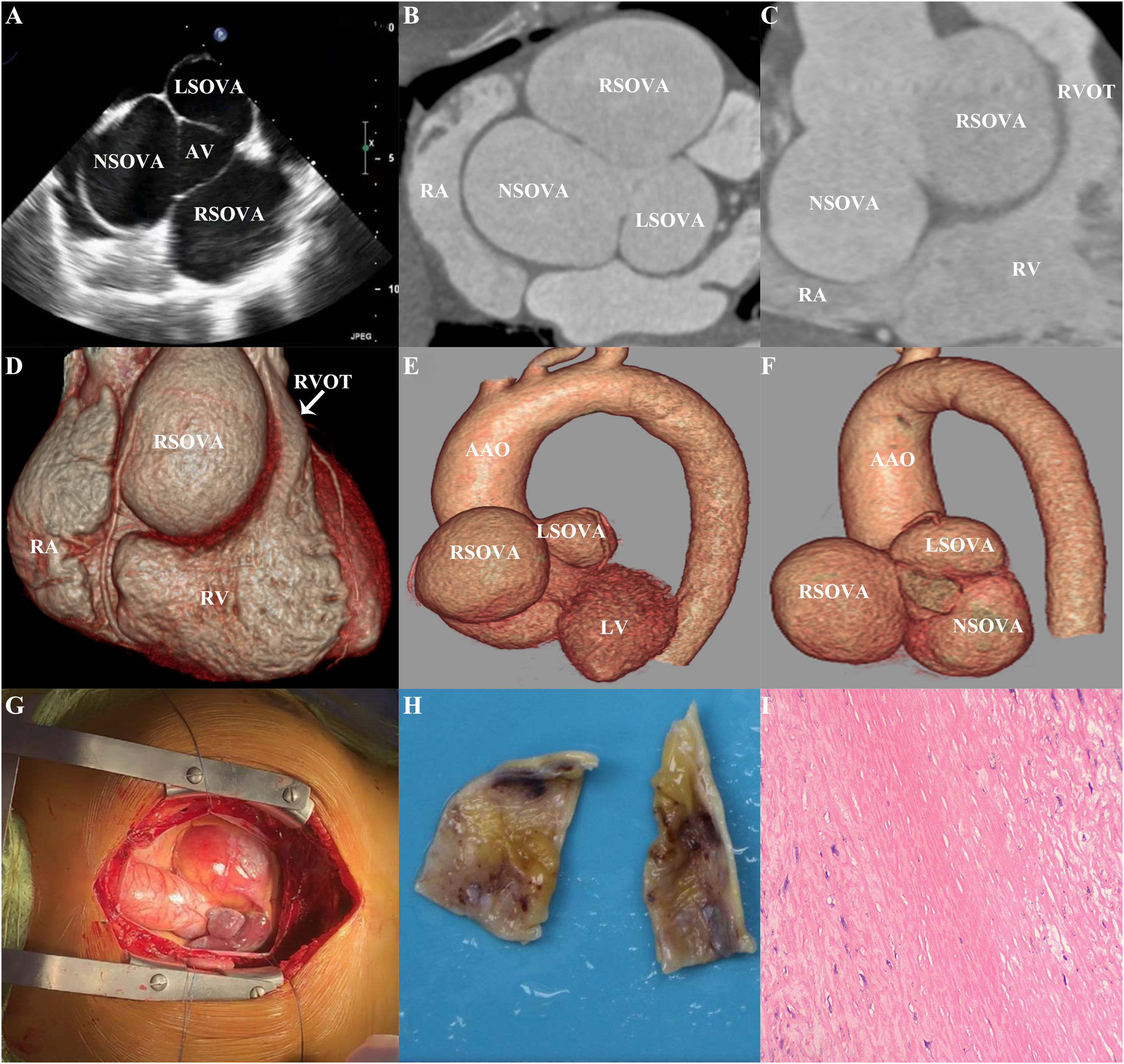
(A) Transthoracic echocardiography shows a giant right sinus of Valsalva aneurysm (SOVA) and noncoronary SOVA. (B and C) Cardiac computerized tomography angiography (CCTA) revealed the SOVA was compressing the bilateral atria and the right ventricle, causing significant right ventricular outflow tract obstruction. (D–F) Three-dimensional volumetric CCTA revealed normal origins of the coronary arteries without substantial compression or stenosis. (G) During the operation, the giant RSOVA and NSOVA were observed with a maximum diameter of 96 mm. (H and I) Pathologic examination demonstrated aneurysmal wall fibrosis, fat deposition, and mid-layer necrosis. AV: aortic valve; LA: left atrium; LAD: left anterior descending; NSOVA: noncoronary SOVA; RA: right atrium; RCA: right coronary artery; RSOVA: right sinus of Valsalva aneurysm; RV: right ventricle; RVOT: right ventricular outflow tract.
Sinus of Valsalva aneurysm is a rare cardiac anomaly that can be congenital or acquired. Unruptured aneurysms may be asymptomatic but can present as malignant arrhythmias, acute chest pain, and even sudden cardiac death. Both ruptured and unruptured SOVAs may have fatal complications; thus, prompt diagnosis and surgery are critical.1 TTE can help assess the SOVA, which can cause hemodynamic changes, aortic valve insufficiency, and affect cardiac function.2 CCTA is preferred for evaluating the size, shape, and relation of the aneurysm to cardiac chambers and coronary arteries.3 Surgical correction is the mainstay of treatment.
Ethical approvalThe Institutional Review Board of Wuhan Asia Heart Hospital approved this study and the patient's informed consent was waived (IRB number: SOP-LLWYH-025-03-R1).
FundingThis work was funded by the Wuhan Clinical Medical Research Center for Cardiovascular Imaging (CMRC202307); Scientific Research Projects from Wuhan Municipal Health Commission (WX23Q44).
Conflict of interestAll authors declare that there is no conflict of interest.